6ZTW | pdb_00006ztw

Crystal Structure of catalase HPII from Escherichia coli (serendipitously crystallized)


Experimental Data Snapshot

  • Method: X-RAY DIFFRACTION
  • Resolution: 1.84 Å
  • R-Value Free: 
    0.184 (Depositor), 0.184 (DCC) 
  • R-Value Work: 
    0.144 (Depositor), 0.145 (DCC) 
  • R-Value Observed: 
    0.144 (Depositor) 

Starting Model: experimental
View more details

wwPDB Validation 3D Report Full Report

Validation slider image for 6ZTW

Ligand Structure Quality Assessment 


This is version 1.5 of the entry. See complete history

Literature

Serendipitous crystallization of E. coli HPII catalase, a sequel to "the tale usually not told".

Grzechowiak, M.Sekula, B.Jaskolski, M.Ruszkowski, M.

(2021) Acta Biochim Pol 68: 29-31

  • DOI: https://doi.org/10.18388/abp.2020_5501
  • Primary Citation Related Structures: 
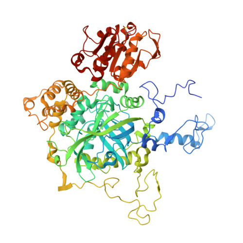
    6ZTV, 6ZTW, 6ZTX

  • PubMed Abstract: 

    Protein crystallographers are well aware of the trap of crystallizing E. coli proteins instead of the macromolecule of interest if heterologous recombinant protein expression in E. coli was part of the experimental pipeline. Among the well-known culprits are YodA metal-binding lipocalin (25 kDa) and YadF carbonic anhydrase (a tetramer of 25 kDa subunits). We report a novel crystal form of another such culprit, E. coli HPII catalase, which is a tetrameric protein of ~340 kDa molecular weight. HPII is likely to contaminate recombinant protein samples, co-purify, and then co-crystallize with the target proteins, especially if their masses in size exclusion chromatography are ~300-400 kDa. What makes this case more interesting but also parlous, is the fact that HPII can crystallize from very low concentrations, even well below 1 mg/mL.


  • Organizational Affiliation
    • Institute of Bioorganic Chemistry, Polish Academy of Sciences, Poznań, Poland.

Macromolecule Content 

  • Total Structure Weight: 682.04 kDa 
  • Atom Count: 51,313 
  • Modeled Residue Count: 5,809 
  • Deposited Residue Count: 6,024 
  • Unique protein chains: 1

Macromolecules

Find similar proteins by:|  3D Structure
Entity ID: 1
MoleculeChains  Sequence LengthOrganismDetailsImage
Catalase HPII
A, B, C, D, E
A, B, C, D, E, F, G, H
753Escherichia coli K-12Mutation(s): 1 
EC: 1.11.1.6
UniProt
Find proteins for P21179 (Escherichia coli (strain K12))
Explore P21179 
Go to UniProtKB:  P21179
Entity Groups
Sequence Clusters30% Identity50% Identity70% Identity90% Identity95% Identity100% Identity
UniProt GroupP21179
Sequence Annotations
Expand
Reference Sequence

Small Molecules

Ligands 5 Unique
IDChains Name / Formula / InChI Key2D Diagram3D Interactions
HDD
(Subject of Investigation/LOI)

Query on HDD


Download:Ideal Coordinates CCD File
EA [auth F]
I [auth A]
IA [auth G]
LA [auth H]
M [auth B]
EA [auth F],
I [auth A],
IA [auth G],
LA [auth H],
M [auth B],
S [auth C],
X [auth D],
Z [auth E]
CIS-HEME D HYDROXYCHLORIN GAMMA-SPIROLACTONE
C34 H32 Fe N4 O5
UMGOPAWIGKFTRK-QQDQPIDJSA-N
TRS

Query on TRS


Download:Ideal Coordinates CCD File
AA [auth E]
BA [auth E]
FA [auth F]
J [auth A]
JA [auth G]
AA [auth E],
BA [auth E],
FA [auth F],
J [auth A],
JA [auth G],
MA [auth H],
N [auth B],
T [auth C],
Y [auth D]
2-AMINO-2-HYDROXYMETHYL-PROPANE-1,3-DIOL
C4 H12 N O3
LENZDBCJOHFCAS-UHFFFAOYSA-O
MPD

Query on MPD


Download:Ideal Coordinates CCD File
O [auth B](4S)-2-METHYL-2,4-PENTANEDIOL
C6 H14 O2
SVTBMSDMJJWYQN-YFKPBYRVSA-N
GOL

Query on GOL


Download:Ideal Coordinates CCD File
DA [auth E]
K [auth A]
KA [auth G]
L [auth A]
R [auth B]
DA [auth E],
K [auth A],
KA [auth G],
L [auth A],
R [auth B],
W [auth C]
GLYCEROL
C3 H8 O3
PEDCQBHIVMGVHV-UHFFFAOYSA-N
EDO

Query on EDO


Download:Ideal Coordinates CCD File
CA [auth E]
GA [auth F]
HA [auth F]
P [auth B]
Q [auth B]
CA [auth E],
GA [auth F],
HA [auth F],
P [auth B],
Q [auth B],
U [auth C],
V [auth C]
1,2-ETHANEDIOL
C2 H6 O2
LYCAIKOWRPUZTN-UHFFFAOYSA-N
Modified Residues  1 Unique
IDChains TypeFormula2D DiagramParent
OCS
Query on OCS
A, B, C, D, E
A, B, C, D, E, F, G, H
L-PEPTIDE LINKINGC3 H7 N O5 SCYS

Experimental Data & Validation

Experimental Data

  • Method: X-RAY DIFFRACTION
  • Resolution: 1.84 Å
  • R-Value Free:  0.184 (Depositor), 0.184 (DCC) 
  • R-Value Work:  0.144 (Depositor), 0.145 (DCC) 
  • R-Value Observed: 0.144 (Depositor) 
Space Group: P 1 21 1
Unit Cell:
Length ( Å )Angle ( ˚ )
a = 121.188α = 90
b = 168.071β = 105.239
c = 137.982γ = 90
Software Package:
Software NamePurpose
PHENIXrefinement
XDSdata reduction
XSCALEdata scaling
PHASERphasing

Structure Validation

View Full Validation Report


Ligand Structure Quality Assessment 


Entry History 

& Funding Information

Deposition Data


Funding OrganizationLocationGrant Number
Polish National Science CentrePolandSONATA 2018/31/D/NZ1/03630
Polish National Science CentrePolandSYMFONIA 2016/20/W/ST5/00478
National Institutes of Health/National Cancer Institute (NIH/NCI)United StatesIntramural Research Program

Revision History  (Full details and data files)

  • Version 1.0: 2020-10-07
    Type: Initial release
  • Version 1.1: 2021-02-10
    Changes: Database references
  • Version 1.2: 2021-02-24
    Changes: Database references
  • Version 1.3: 2021-03-03
    Changes: Database references
  • Version 1.4: 2021-03-31
    Changes: Database references
  • Version 1.5: 2024-01-31
    Changes: Data collection, Database references, Refinement description