6WV6

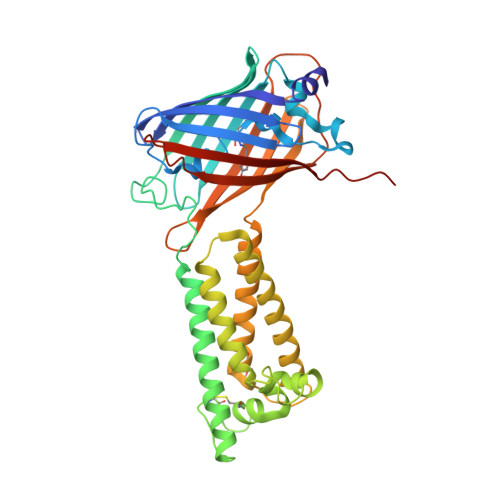
Human VKOR with phenindione


Experimental Data Snapshot

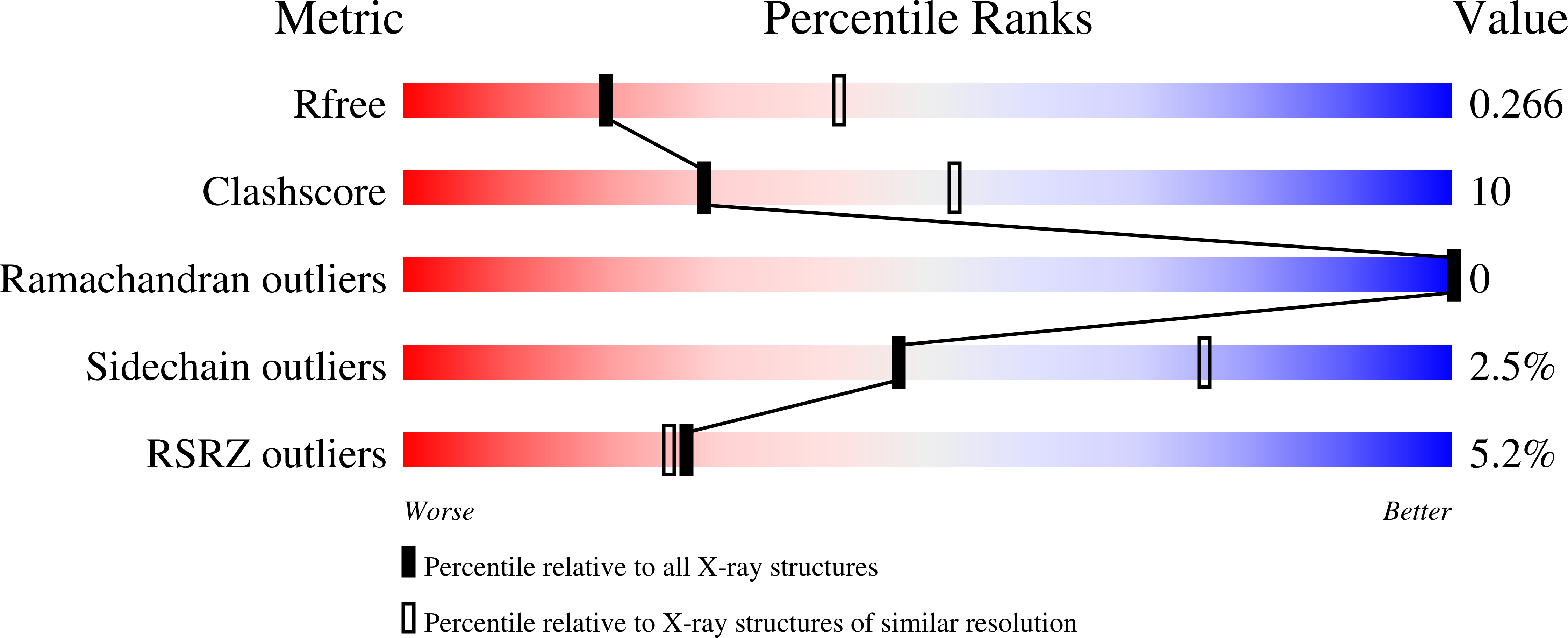
  • Method: X-RAY DIFFRACTION
  • Resolution: 2.70 Å
  • R-Value Free: 0.268 
  • R-Value Work: 0.226 
  • R-Value Observed: 0.228 

Starting Model: experimental
View more details

wwPDB Validation   3D Report Full Report


Ligand Structure Quality Assessment 


This is version 1.4 of the entry. See complete history


Literature

Structural basis of antagonizing the vitamin K catalytic cycle for anticoagulation.

Liu, S.Li, S.Shen, G.Sukumar, N.Krezel, A.M.Li, W.

(2021) Science 371

  • DOI: https://doi.org/10.1126/science.abc5667
  • Primary Citation of Related Structures:  
    6WV3, 6WV4, 6WV5, 6WV6, 6WV7, 6WV8, 6WV9, 6WVA, 6WVB, 6WVH, 6WVI

  • PubMed Abstract: 

    Vitamin K antagonists are widely used anticoagulants that target vitamin K epoxide reductases (VKOR), a family of integral membrane enzymes. To elucidate their catalytic cycle and inhibitory mechanism, we report 11 x-ray crystal structures of human VKOR and pufferfish VKOR-like, with substrates and antagonists in different redox states. Substrates entering the active site in a partially oxidized state form cysteine adducts that induce an open-to-closed conformational change, triggering reduction. Binding and catalysis are facilitated by hydrogen-bonding interactions in a hydrophobic pocket. The antagonists bind specifically to the same hydrogen-bonding residues and induce a similar closed conformation. Thus, vitamin K antagonists act through mimicking the key interactions and conformational changes required for the VKOR catalytic cycle.


  • Organizational Affiliation

    Department of Biochemistry and Molecular Biophysics, Washington University School of Medicine, St. Louis, MO 63110, USA.


Macromolecules
Find similar proteins by:  (by identity cutoff)  |  3D Structure
Entity ID: 1
MoleculeChains Sequence LengthOrganismDetailsImage
Vitamin K epoxide reductase, termini restrained by green fluorescent protein390Aequorea victoriaHomo sapiensMutation(s): 1 
Gene Names: gfpVKORC1VKORMSTP134MSTP576UNQ308/PRO351
EC: 1.17.4.4
UniProt & NIH Common Fund Data Resources
Find proteins for Q9BQB6 (Homo sapiens)
Go to UniProtKB:  Q9BQB6
PHAROS:  Q9BQB6
GTEx:  ENSG00000167397 
Find proteins for P42212 (Aequorea victoria)
Go to UniProtKB:  P42212
Sequence Annotations
Expand
  • Reference Sequence
Small Molecules
Modified Residues  1 Unique
IDChains TypeFormula2D DiagramParent
CRO
Query on CRO
A
L-PEPTIDE LINKINGC15 H17 N3 O5THR, TYR, GLY
Experimental Data & Validation

Experimental Data

  • Method: X-RAY DIFFRACTION
  • Resolution: 2.70 Å
  • R-Value Free: 0.268 
  • R-Value Work: 0.226 
  • R-Value Observed: 0.228 
  • Space Group: P 21 21 2
Unit Cell:
Length ( Å )Angle ( ˚ )
a = 155.019α = 90
b = 84.878β = 90
c = 42.558γ = 90
Software Package:
Software NamePurpose
PHENIXrefinement
HKL-2000data scaling
PDB_EXTRACTdata extraction
HKL-2000data reduction
PHASERphasing

Structure Validation

View Full Validation Report


Ligand Structure Quality Assessment 


Entry History & Funding Information

Deposition Data


Funding OrganizationLocationGrant Number
National Institutes of Health/National Heart, Lung, and Blood Institute (NIH/NHLBI)United StatesR01 HL121718
Other privateUnited StatesForefront of Science Award
Other privateUnited StatesMCII 2020-854
National Institutes of Health/National Eye Institute (NIH/NEI)United StatesR21 EY028705
National Institutes of Health/National Institute of General Medical Sciences (NIH/NIGMS)United StatesR01 GM131008
National Institutes of Health/National Institute of General Medical Sciences (NIH/NIGMS)United StatesP30 GM124165

Revision History  (Full details and data files)

  • Version 1.0: 2020-11-11
    Type: Initial release
  • Version 1.1: 2020-11-25
    Changes: Database references
  • Version 1.2: 2021-01-13
    Changes: Database references
  • Version 1.3: 2023-10-18
    Changes: Data collection, Database references, Refinement description
  • Version 1.4: 2023-11-15
    Changes: Data collection