6TXK

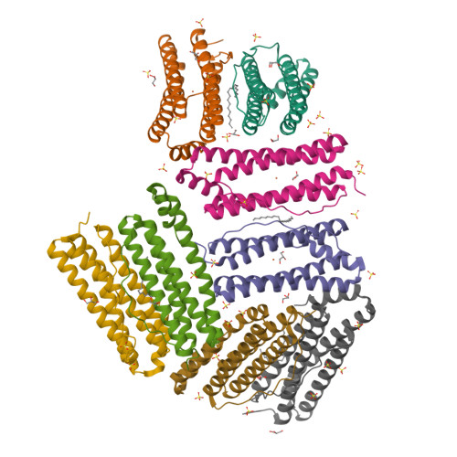
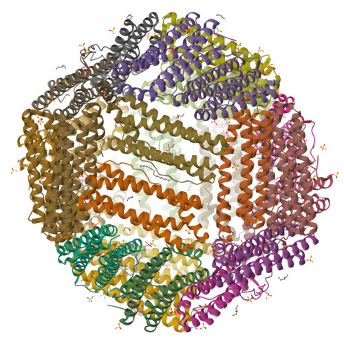
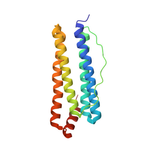
Crystal structure of thermotoga maritima E65K Ferritin


Experimental Data Snapshot

  • Method: X-RAY DIFFRACTION
  • Resolution: 2.36 Å
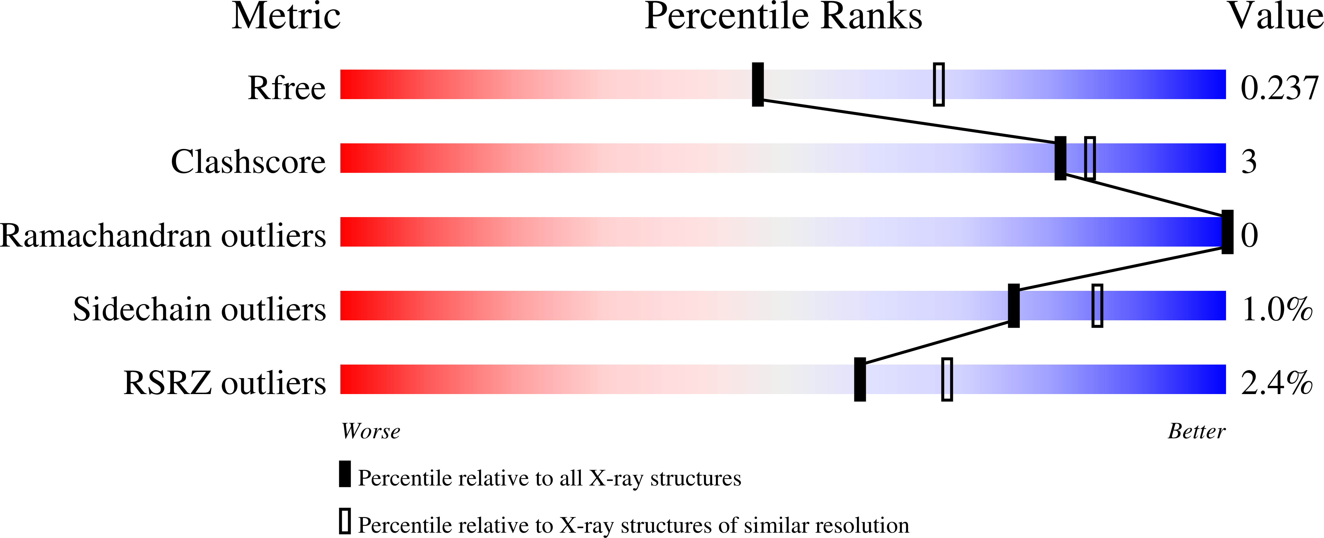
  • R-Value Free: 0.236 
  • R-Value Work: 0.201 
  • R-Value Observed: 0.202 

Starting Model: experimental
View more details

wwPDB Validation   3D Report Full Report


Ligand Structure Quality Assessment 


This is version 1.2 of the entry. See complete history


Literature

A single residue can modulate nanocage assembly in salt dependent ferritin.

Kumar, M.Markiewicz-Mizera, J.Janna Olmos, J.D.Wilk, P.Grudnik, P.Biela, A.P.Jemiola-Rzeminska, M.Gorecki, A.Chakraborti, S.Heddle, J.G.

(2021) Nanoscale 13: 11932-11942

  • DOI: https://doi.org/10.1039/d1nr01632f
  • Primary Citation of Related Structures:  
    6TXH, 6TXI, 6TXJ, 6TXK, 6TXL, 6TXM, 6TXN

  • PubMed Abstract: 

    Cage forming proteins have numerous potential applications in biomedicine and biotechnology, where the iron storage ferritin is a widely used example. However, controlling ferritin cage assembly/disassembly remains challenging, typically requiring extreme conditions incompatible with many desirable cargoes, particularly for more fragile biopharmaceuticals. Recently, a ferritin from the hyperthermophile bacterium Thermotoga maritima (TmFtn) has been shown to have reversible assembly under mild conditions, offering greater potential biocompatibility in terms of cargo access and encapsulation. Like Archeoglobus fulgidus ferritin (AfFtn), TmFtn forms 24mer cages mediated by metal ions (Mg2+). We have solved the crystal structure of the wild type TmFtn and several mutants displaying different assembly/disassembly properties. These data combined with other biophysical studies allow us to suggest candidate interfacial amino acids crucial in controlling assembly. This work deepens our understanding of how these ferritin complexes assemble and is a useful step towards production of triggerable ferritins in which these properties can be finely designed and controlled.


  • Organizational Affiliation

    Malopolska Centre of Biotechnology, Jagiellonian University, Gronostajowa 7A, 30-392 Krakow, Poland. soumyabiochem@gmail.com Jonathan.heddle@uj.edu.pl.


Macromolecules
Find similar proteins by:  (by identity cutoff)  |  3D Structure
Entity ID: 1
MoleculeChains Sequence LengthOrganismDetailsImage
Ferritin
A, B, C, D, E
A, B, C, D, E, F, G, H
164Thermotoga maritima MSB8Mutation(s): 1 
Gene Names: TM_1128
EC: 1.16.3.2
UniProt
Find proteins for Q9X0L2 (Thermotoga maritima (strain ATCC 43589 / DSM 3109 / JCM 10099 / NBRC 100826 / MSB8))
Explore Q9X0L2 
Go to UniProtKB:  Q9X0L2
Entity Groups  
Sequence Clusters30% Identity50% Identity70% Identity90% Identity95% Identity100% Identity
UniProt GroupQ9X0L2
Sequence Annotations
Expand
  • Reference Sequence
Small Molecules
Ligands 4 Unique
IDChains Name / Formula / InChI Key2D Diagram3D Interactions
LFA
Query on LFA

Download Ideal Coordinates CCD File 
DA [auth C],
IB [auth H],
TA [auth E],
W [auth B]
EICOSANE
C20 H42
CBFCDTFDPHXCNY-UHFFFAOYSA-N
SO4
Query on SO4

Download Ideal Coordinates CCD File 
BB [auth G]
CB [auth G]
DB [auth G]
EA [auth C]
EB [auth G]
BB [auth G],
CB [auth G],
DB [auth G],
EA [auth C],
EB [auth G],
FA [auth C],
GA [auth C],
GB [auth H],
IA [auth D],
J [auth A],
JB [auth H],
KB [auth H],
L [auth A],
LA [auth D],
LB [auth H],
M [auth A],
MA [auth D],
N [auth A],
NA [auth D],
O [auth A],
OA [auth D],
PA [auth D],
Q [auth B],
R [auth B],
RA [auth E],
T [auth B],
UA [auth E],
WA [auth F],
X [auth B],
Y [auth B],
YA [auth F],
Z [auth B]
SULFATE ION
O4 S
QAOWNCQODCNURD-UHFFFAOYSA-L
GOL
Query on GOL

Download Ideal Coordinates CCD File 
AB [auth G]
BA [auth C]
CA [auth C]
HB [auth H]
JA [auth D]
AB [auth G],
BA [auth C],
CA [auth C],
HB [auth H],
JA [auth D],
K [auth A],
KA [auth D],
S [auth B],
SA [auth E],
U [auth B],
V [auth B],
XA [auth F]
GLYCEROL
C3 H8 O3
PEDCQBHIVMGVHV-UHFFFAOYSA-N
FE
Query on FE

Download Ideal Coordinates CCD File 
AA [auth C]
FB [auth H]
HA [auth D]
I [auth A]
P [auth B]
AA [auth C],
FB [auth H],
HA [auth D],
I [auth A],
P [auth B],
QA [auth E],
VA [auth F],
ZA [auth G]
FE (III) ION
Fe
VTLYFUHAOXGGBS-UHFFFAOYSA-N
Experimental Data & Validation

Experimental Data

  • Method: X-RAY DIFFRACTION
  • Resolution: 2.36 Å
  • R-Value Free: 0.236 
  • R-Value Work: 0.201 
  • R-Value Observed: 0.202 
  • Space Group: H 3 2
Unit Cell:
Length ( Å )Angle ( ˚ )
a = 175.811α = 90
b = 175.811β = 90
c = 354.136γ = 120
Software Package:
Software NamePurpose
XSCALEdata scaling
PHASERphasing
PHENIXrefinement
PDB_EXTRACTdata extraction
XDSdata reduction

Structure Validation

View Full Validation Report


Ligand Structure Quality Assessment 


Entry History & Funding Information

Deposition Data


Funding OrganizationLocationGrant Number
Foundation for Polish SciencePolandHoming/2017-3/22

Revision History  (Full details and data files)

  • Version 1.0: 2021-07-28
    Type: Initial release
  • Version 1.1: 2022-08-10
    Changes: Database references
  • Version 1.2: 2024-01-24
    Changes: Data collection, Database references, Refinement description