6TTM | pdb_00006ttm

Hyoscyamine 6-hydroxylase in complex with N-oxalylglycine and hyoscyamine


Experimental Data Snapshot

  • Method: X-RAY DIFFRACTION
  • Resolution: 1.91 Å
  • R-Value Free: 
    0.200 (Depositor), 0.203 (DCC) 
  • R-Value Work: 
    0.159 (Depositor), 0.163 (DCC) 
  • R-Value Observed: 
    0.161 (Depositor) 

Starting Model: experimental
View more details

wwPDB Validation 3D Report Full Report

Validation slider image for 6TTM

Ligand Structure Quality Assessment 


This is version 1.3 of the entry. See complete history

Literature

Regioselectivity of hyoscyamine 6 beta-hydroxylase-catalysed hydroxylation as revealed by high-resolution structural information and QM/MM calculations.

Kluza, A.Wojdyla, Z.Mrugala, B.Kurpiewska, K.Porebski, P.J.Niedzialkowska, E.Minor, W.Weiss, M.S.Borowski, T.

(2020) Dalton Trans 49: 4454-4469

  • DOI: https://doi.org/10.1039/d0dt00302f
  • Primary Citation Related Structures: 
    6TTM, 6TTN, 6TTO

  • PubMed Abstract: 

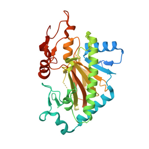
    Hyoscyamine 6β-hydroxylase (H6H) is a bifunctional non-heme 2-oxoglutarate/Fe2+-dependent dioxygenase that catalyzes the two final steps in the biosynthesis of scopolamine. Based on high resolution crystal structures of H6H from Datura metel, detailed information on substrate binding was obtained that provided insights into the onset of the enzymatic process. In particular, the role of two prominent residues was revealed - Glu-116 that interacts with the tertiary amine located on the hyoscyamine tropane moiety and Tyr-326 that forms CH-π hydrogen bonds with the hyoscyamine phenyl ring. The structures were used as the basis for QM/MM calculations that provided an explanation for the regioselectivity of the hydroxylation reaction on the hyoscyamine tropane moiety (C6 vs. C7) and quantified contributions of active site residues to respective barrier heights.


  • Organizational Affiliation
    • Jerzy Haber Institute of Catalysis and Surface Chemistry, Polish Academy of Sciences, Niezapominajek 8, PL-30239 Krakow, Poland. ncborows@cyf-kr.edu.pl.

Macromolecule Content 

  • Total Structure Weight: 40.68 kDa 
  • Atom Count: 2,679 
  • Modeled Residue Count: 299 
  • Deposited Residue Count: 350 
  • Unique protein chains: 1

Macromolecules

Find similar proteins by:|  3D Structure
Entity ID: 1
MoleculeChains  Sequence LengthOrganismDetailsImage
Hyoscyamine 6 beta-hydroxylase350Datura metelMutation(s): 0 
Gene Names: H6H
UniProt
Find proteins for Q6EZB3 (Datura metel)
Explore Q6EZB3 
Go to UniProtKB:  Q6EZB3
Entity Groups
Sequence Clusters30% Identity50% Identity70% Identity90% Identity95% Identity100% Identity
UniProt GroupQ6EZB3
Sequence Annotations
Expand
Reference Sequence

Small Molecules

Ligands 7 Unique
IDChains Name / Formula / InChI Key2D Diagram3D Interactions
HYO
(Subject of Investigation/LOI)

Query on HYO


Download:Ideal Coordinates CCD File
C [auth A][(1S,5R)-8-methyl-8-azabicyclo[3.2.1]octan-3-yl] (2S)-3-hydroxy-2-phenylpropanoate
C17 H23 N O3
RKUNBYITZUJHSG-FXUDXRNXSA-N
OGA
(Subject of Investigation/LOI)

Query on OGA


Download:Ideal Coordinates CCD File
E [auth A]N-OXALYLGLYCINE
C4 H5 N O5
BIMZLRFONYSTPT-UHFFFAOYSA-N
PEG

Query on PEG


Download:Ideal Coordinates CCD File
B [auth A]DI(HYDROXYETHYL)ETHER
C4 H10 O3
MTHSVFCYNBDYFN-UHFFFAOYSA-N
SR

Query on SR


Download:Ideal Coordinates CCD File
H [auth A]STRONTIUM ION
Sr
PWYYWQHXAPXYMF-UHFFFAOYSA-N
EDO

Query on EDO


Download:Ideal Coordinates CCD File
F [auth A],
G [auth A]
1,2-ETHANEDIOL
C2 H6 O2
LYCAIKOWRPUZTN-UHFFFAOYSA-N
NI

Query on NI


Download:Ideal Coordinates CCD File
D [auth A]NICKEL (II) ION
Ni
VEQPNABPJHWNSG-UHFFFAOYSA-N
UNX

Query on UNX


Download:Ideal Coordinates CCD File
I [auth A],
J [auth A]
UNKNOWN ATOM OR ION
X

Experimental Data & Validation

Experimental Data

  • Method: X-RAY DIFFRACTION
  • Resolution: 1.91 Å
  • R-Value Free:  0.200 (Depositor), 0.203 (DCC) 
  • R-Value Work:  0.159 (Depositor), 0.163 (DCC) 
  • R-Value Observed: 0.161 (Depositor) 
Space Group: P 21 21 21
Unit Cell:
Length ( Å )Angle ( ˚ )
a = 68.536α = 90
b = 70.78β = 90
c = 78.63γ = 90
Software Package:
Software NamePurpose
PHENIXrefinement
REFMACrefinement
PDB_EXTRACTdata extraction
HKL-2000data reduction
HKL-2000data scaling
MoRDaphasing

Structure Validation

View Full Validation Report


Ligand Structure Quality Assessment 


Entry History 

& Funding Information

Deposition Data


Funding OrganizationLocationGrant Number
Polish National Science CentrePoland2014/14/E/NZ1/00053

Revision History  (Full details and data files)

  • Version 1.0: 2020-03-18
    Type: Initial release
  • Version 1.1: 2020-03-25
    Changes: Database references
  • Version 1.2: 2020-04-15
    Changes: Database references
  • Version 1.3: 2024-01-24
    Changes: Data collection, Database references, Derived calculations, Refinement description