6TQU

The crystal structure of the MSP domain of human MOSPD2 in complex with the Phospho-FFAT motif of STARD3.


Experimental Data Snapshot

  • Method: X-RAY DIFFRACTION
  • Resolution: 2.40 Å
  • R-Value Free: 0.240 
  • R-Value Work: 0.183 
  • R-Value Observed: 0.186 

Starting Model: experimental
View more details

wwPDB Validation   3D Report Full Report


This is version 1.2 of the entry. See complete history


Literature

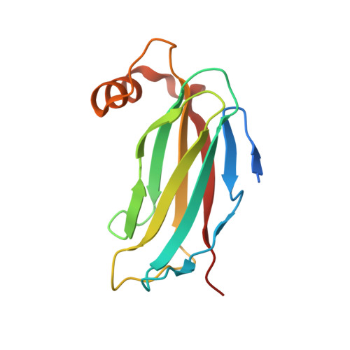
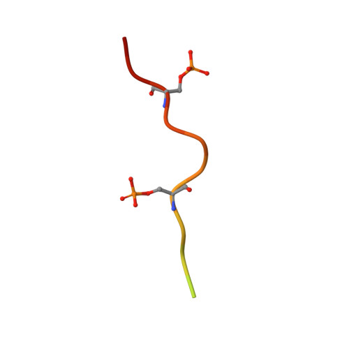
FFAT motif phosphorylation controls formation and lipid transfer function of inter-organelle contacts.

Di Mattia, T.Martinet, A.Ikhlef, S.McEwen, A.G.Nomine, Y.Wendling, C.Poussin-Courmontagne, P.Voilquin, L.Eberling, P.Ruffenach, F.Cavarelli, J.Slee, J.Levine, T.P.Drin, G.Tomasetto, C.Alpy, F.

(2020) EMBO J 39: e104369-e104369

  • DOI: https://doi.org/10.15252/embj.2019104369
  • Primary Citation of Related Structures:  
    6TQR, 6TQS, 6TQT, 6TQU

  • PubMed Abstract: 

    Organelles are physically connected in membrane contact sites. The endoplasmic reticulum possesses three major receptors, VAP-A, VAP-B, and MOSPD2, which interact with proteins at the surface of other organelles to build contacts. VAP-A, VAP-B, and MOSPD2 contain an MSP domain, which binds a motif named FFAT (two phenylalanines in an acidic tract). In this study, we identified a non-conventional FFAT motif where a conserved acidic residue is replaced by a serine/threonine. We show that phosphorylation of this serine/threonine is critical for non-conventional FFAT motifs (named Phospho-FFAT) to be recognized by the MSP domain. Moreover, structural analyses of the MSP domain alone or in complex with conventional and Phospho-FFAT peptides revealed new mechanisms of interaction. Based on these new insights, we produced a novel prediction algorithm, which expands the repertoire of candidate proteins with a Phospho-FFAT that are able to create membrane contact sites. Using a prototypical tethering complex made by STARD3 and VAP, we showed that phosphorylation is instrumental for the formation of ER-endosome contacts, and their sterol transfer function. This study reveals that phosphorylation acts as a general switch for inter-organelle contacts.


  • Organizational Affiliation

    Institut de Génétique et de Biologie Moléculaire et Cellulaire (IGBMC), Illkirch, France.


Macromolecules
Find similar proteins by:  (by identity cutoff)  |  3D Structure
Entity ID: 1
MoleculeChains Sequence LengthOrganismDetailsImage
Motile sperm domain-containing protein 2
A, B
135Homo sapiensMutation(s): 0 
Gene Names: MOSPD2
UniProt & NIH Common Fund Data Resources
Find proteins for Q8NHP6 (Homo sapiens)
Explore Q8NHP6 
Go to UniProtKB:  Q8NHP6
PHAROS:  Q8NHP6
GTEx:  ENSG00000130150 
Entity Groups  
Sequence Clusters30% Identity50% Identity70% Identity90% Identity95% Identity100% Identity
UniProt GroupQ8NHP6
Sequence Annotations
Expand
  • Reference Sequence

Find similar proteins by:  Sequence   |   3D Structure  

Entity ID: 2
MoleculeChains Sequence LengthOrganismDetailsImage
StAR-related lipid transfer protein 3
C, D
21Homo sapiensMutation(s): 0 
UniProt & NIH Common Fund Data Resources
Find proteins for Q14849 (Homo sapiens)
Explore Q14849 
Go to UniProtKB:  Q14849
GTEx:  ENSG00000131748 
Entity Groups  
Sequence Clusters30% Identity50% Identity70% Identity90% Identity95% Identity100% Identity
UniProt GroupQ14849
Sequence Annotations
Expand
  • Reference Sequence
Small Molecules
Ligands 1 Unique
IDChains Name / Formula / InChI Key2D Diagram3D Interactions
SO4
Query on SO4

Download Ideal Coordinates CCD File 
E [auth B]SULFATE ION
O4 S
QAOWNCQODCNURD-UHFFFAOYSA-L
Modified Residues  1 Unique
IDChains TypeFormula2D DiagramParent
SEP
Query on SEP
C, D
L-PEPTIDE LINKINGC3 H8 N O6 PSER
Experimental Data & Validation

Experimental Data

  • Method: X-RAY DIFFRACTION
  • Resolution: 2.40 Å
  • R-Value Free: 0.240 
  • R-Value Work: 0.183 
  • R-Value Observed: 0.186 
  • Space Group: P 41 21 2
Unit Cell:
Length ( Å )Angle ( ˚ )
a = 87.968α = 90
b = 87.968β = 90
c = 95.13γ = 90
Software Package:
Software NamePurpose
XDSdata reduction
Aimlessdata scaling
PHASERphasing
PHENIXrefinement
PDB_EXTRACTdata extraction

Structure Validation

View Full Validation Report


Entry History & Funding Information

Deposition Data


Funding OrganizationLocationGrant Number
French National Research AgencyFranceANR-19-CE44-0003
French National Research AgencyFranceANR-10-INSB-05-01
French National Research AgencyFranceANR-10-LABX-0030-INRT
French National Research AgencyFranceANR-10-IDEX-0002-02.

Revision History  (Full details and data files)

  • Version 1.0: 2020-11-18
    Type: Initial release
  • Version 1.1: 2020-12-16
    Changes: Database references
  • Version 1.2: 2024-01-24
    Changes: Data collection, Database references, Refinement description