6SEK | pdb_00006sek

Crystal Structure of Ancestral Flavin-containing monooxygenase (FMO) 5


Experimental Data Snapshot

  • Method: X-RAY DIFFRACTION
  • Resolution: 2.70 Å
  • R-Value Free: 
    0.295 (Depositor), 0.297 (DCC) 
  • R-Value Work: 
    0.222 (Depositor), 0.227 (DCC) 
  • R-Value Observed: 
    0.226 (Depositor) 

wwPDB Validation 3D Report Full Report

Validation slider image for 6SEK

Ligand Structure Quality Assessment 


This is version 1.4 of the entry. See complete history

Literature

Ancestral-sequence reconstruction unveils the structural basis of function in mammalian FMOs.

Nicoll, C.R.Bailleul, G.Fiorentini, F.Mascotti, M.L.Fraaije, M.W.Mattevi, A.

(2020) Nat Struct Mol Biol 27: 14-24

  • DOI: https://doi.org/10.1038/s41594-019-0347-2
  • Primary Citation Related Structures: 
    6SE3, 6SEK, 6SEM, 6SF0

  • PubMed Abstract: 

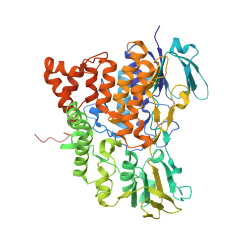
    Flavin-containing monooxygenases (FMOs) are ubiquitous in all domains of life and metabolize a myriad of xenobiotics, including toxins, pesticides and drugs. However, despite their pharmacological importance, structural information remains bereft. To further our understanding behind their biochemistry and diversity, we used ancestral-sequence reconstruction, kinetic and crystallographic techniques to scrutinize three ancient mammalian FMOs: AncFMO2, AncFMO3-6 and AncFMO5. Remarkably, all AncFMOs could be crystallized and were structurally resolved between 2.7- and 3.2-Å resolution. These crystal structures depict the unprecedented topology of mammalian FMOs. Each employs extensive membrane-binding features and intricate substrate-profiling tunnel networks through a conspicuous membrane-adhering insertion. Furthermore, a glutamate-histidine switch is speculated to induce the distinctive Baeyer-Villiger oxidation activity of FMO5. The AncFMOs exhibited catalysis akin to human FMOs and, with sequence identities between 82% and 92%, represent excellent models. Our study demonstrates the power of ancestral-sequence reconstruction as a strategy for the crystallization of proteins.


  • Organizational Affiliation
    • Department of Biology and Biotechnology "Lazzaro Spallanzani", University of Pavia, Pavia, Italy.

Macromolecule Content 

  • Total Structure Weight: 125.12 kDa 
  • Atom Count: 8,329 
  • Modeled Residue Count: 1,008 
  • Deposited Residue Count: 1,066 
  • Unique protein chains: 1

Macromolecules

Find similar proteins by:|  3D Structure
Entity ID: 1
MoleculeChains  Sequence LengthOrganismDetailsImage
Ancestral Flavin-containing monooxygenase 5
A, B
533synthetic constructMutation(s): 0 

Small Molecules

Ligands 5 Unique
IDChains Name / Formula / InChI Key2D Diagram3D Interactions
FAD

Query on FAD


Download:Ideal Coordinates CCD File
C [auth A],
I [auth B]
FLAVIN-ADENINE DINUCLEOTIDE
C27 H33 N9 O15 P2
VWWQXMAJTJZDQX-UYBVJOGSSA-N
NAP

Query on NAP


Download:Ideal Coordinates CCD File
D [auth A],
J [auth B]
NADP NICOTINAMIDE-ADENINE-DINUCLEOTIDE PHOSPHATE
C21 H28 N7 O17 P3
XJLXINKUBYWONI-NNYOXOHSSA-N
LMT

Query on LMT


Download:Ideal Coordinates CCD File
E [auth A]DODECYL-BETA-D-MALTOSIDE
C24 H46 O11
NLEBIOOXCVAHBD-QKMCSOCLSA-N
EPE

Query on EPE


Download:Ideal Coordinates CCD File
F [auth A],
K [auth B]
4-(2-HYDROXYETHYL)-1-PIPERAZINE ETHANESULFONIC ACID
C8 H18 N2 O4 S
JKMHFZQWWAIEOD-UHFFFAOYSA-N
GLC

Query on GLC


Download:Ideal Coordinates CCD File
G [auth A],
H [auth A]
alpha-D-glucopyranose
C6 H12 O6
WQZGKKKJIJFFOK-DVKNGEFBSA-N

Experimental Data & Validation

Experimental Data

  • Method: X-RAY DIFFRACTION
  • Resolution: 2.70 Å
  • R-Value Free:  0.295 (Depositor), 0.297 (DCC) 
  • R-Value Work:  0.222 (Depositor), 0.227 (DCC) 
  • R-Value Observed: 0.226 (Depositor) 
Space Group: P 2 21 21
Unit Cell:
Length ( Å )Angle ( ˚ )
a = 96.782α = 90
b = 100.069β = 90
c = 143.152γ = 90
Software Package:
Software NamePurpose
REFMACrefinement
XDSdata reduction
Aimlessdata scaling
PHASERphasing

Structure Validation

View Full Validation Report


Ligand Structure Quality Assessment 


Entry History 

& Funding Information

Deposition Data


Funding OrganizationLocationGrant Number
European Commission722390

Revision History  (Full details and data files)

  • Version 1.0: 2019-12-25
    Type: Initial release
  • Version 1.1: 2020-01-01
    Changes: Database references
  • Version 1.2: 2020-01-22
    Changes: Database references
  • Version 1.3: 2020-07-29
    Type: Remediation
    Reason: Carbohydrate remediation
    Changes: Data collection, Derived calculations, Structure summary
  • Version 1.4: 2024-05-15
    Changes: Data collection, Database references, Structure summary