6RIX | pdb_00006rix

Crystal structure of MchDnaB-1 intein


Experimental Data Snapshot

  • Method: X-RAY DIFFRACTION
  • Resolution: 1.66 Å
  • R-Value Free: 
    0.214 (Depositor), 0.213 (DCC) 
  • R-Value Work: 
    0.159 (Depositor), 0.158 (DCC) 
  • R-Value Observed: 
    0.161 (Depositor) 

Starting Model: experimental
View more details

wwPDB Validation 3D Report Full Report

Validation slider image for 6RIX

This is version 1.2 of the entry. See complete history

Literature

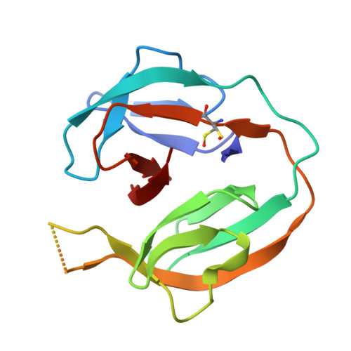
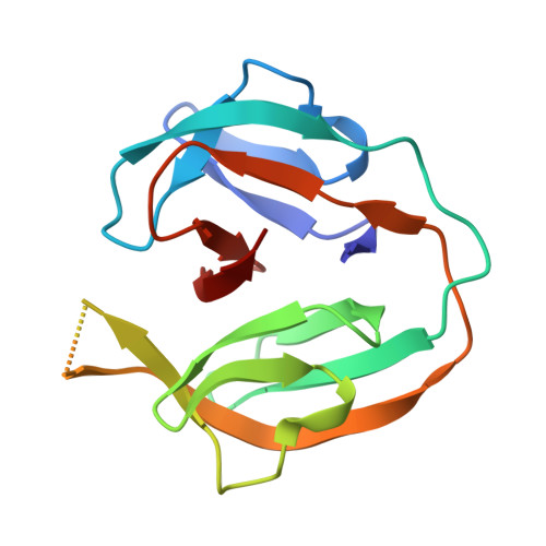
The Convergence of the Hedgehog/Intein Fold in Different Protein Splicing Mechanisms.

Beyer, H.M.Virtanen, S.I.Aranko, A.S.Mikula, K.M.Lountos, G.T.Wlodawer, A.Ollila, O.H.S.Iwai, H.

(2020) Int J Mol Sci 21

  • DOI: https://doi.org/10.3390/ijms21218367
  • Primary Citation Related Structures: 
    6RIX, 6RIY, 6RIZ

  • PubMed Abstract: 

    Protein splicing catalyzed by inteins utilizes many different combinations of amino-acid types at active sites. Inteins have been classified into three classes based on their characteristic sequences. We investigated the structural basis of the protein splicing mechanism of class 3 inteins by determining crystal structures of variants of a class 3 intein from Mycobacterium chimaera and molecular dynamics simulations, which suggested that the class 3 intein utilizes a different splicing mechanism from that of class 1 and 2 inteins. The class 3 intein uses a bond cleavage strategy reminiscent of proteases but share the same Hedgehog/INTein (HINT) fold of other intein classes. Engineering of class 3 inteins from a class 1 intein indicated that a class 3 intein would unlikely evolve directly from a class 1 or 2 intein. The HINT fold appears as structural and functional solution for trans -peptidyl and trans -esterification reactions commonly exploited by diverse mechanisms using different combinations of amino-acid types for the active-site residues.


  • Organizational Affiliation
    • Institute of Biotechnology, University of Helsinki, P.O. Box 65, FIN-00014 Helsinki, Finland.

Macromolecule Content 

  • Total Structure Weight: 30.19 kDa 
  • Atom Count: 2,175 
  • Modeled Residue Count: 261 
  • Deposited Residue Count: 290 
  • Unique protein chains: 2

Macromolecules

Find similar proteins by:|  3D Structure
Entity ID: 1
MoleculeChains  Sequence LengthOrganismDetailsImage
Replicative DNA helicase145Mycobacterium intracellulare subsp. chimaeraMutation(s): 0 
Gene Names: BWK49_00400MYCOZU1_00080
EC: 3.6.4.12
Find similar proteins by:|  3D Structure
Entity ID: 2
MoleculeChains  Sequence LengthOrganismDetailsImage
Replicative DNA helicase145Mycobacterium intracellulare subsp. chimaeraMutation(s): 0 
Gene Names: BWK49_00400MYCOZU1_00080
EC: 3.6.4.12

Small Molecules

Ligands 1 Unique
IDChains Name / Formula / InChI Key2D Diagram3D Interactions
CL

Query on CL


Download:Ideal Coordinates CCD File
C [auth B]CHLORIDE ION
Cl
VEXZGXHMUGYJMC-UHFFFAOYSA-M
Modified Residues  1 Unique
IDChains TypeFormula2D DiagramParent
CSX
Query on CSX
A
L-PEPTIDE LINKINGC3 H7 N O3 SCYS

Experimental Data & Validation

Experimental Data

  • Method: X-RAY DIFFRACTION
  • Resolution: 1.66 Å
  • R-Value Free:  0.214 (Depositor), 0.213 (DCC) 
  • R-Value Work:  0.159 (Depositor), 0.158 (DCC) 
  • R-Value Observed: 0.161 (Depositor) 
Space Group: P 1 21 1
Unit Cell:
Length ( Å )Angle ( ˚ )
a = 31.246α = 90
b = 95.344β = 107.14
c = 35.796γ = 90
Software Package:
Software NamePurpose
PHENIXrefinement
HKL-3000data reduction
HKL-3000data scaling
PHASERphasing
PHENIXmodel building

Structure Validation

View Full Validation Report


Entry History 

& Funding Information

Deposition Data


Funding OrganizationLocationGrant Number
Academy of FinlandFinland137995, 277335
Novo Nordisk FoundationFinlandNNF17OC0025402
Sigrid Juselius FoundationFinland--

Revision History  (Full details and data files)

  • Version 1.0: 2020-05-13
    Type: Initial release
  • Version 1.1: 2020-12-23
    Changes: Database references
  • Version 1.2: 2024-01-24
    Changes: Data collection, Database references, Refinement description