6R3U | pdb_00006r3u

Endo-levanase BT1760 mutant E221A from Bacteroides thetaiotaomicron complexed with levantetraose


Experimental Data Snapshot

  • Method: X-RAY DIFFRACTION
  • Resolution: 1.90 Å
  • R-Value Free: 
    0.190 (Depositor), 0.196 (DCC) 
  • R-Value Work: 
    0.161 (Depositor), 0.166 (DCC) 

Starting Model: experimental
View more details

wwPDB Validation 3D Report Full Report

Validation slider image for 6R3U

This is version 2.1 of the entry. See complete history

Literature

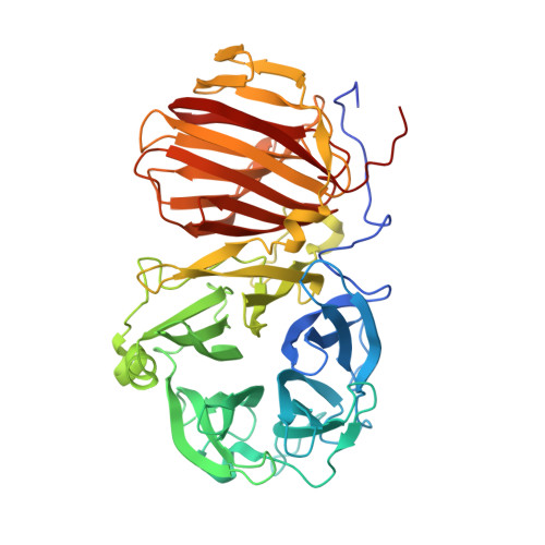
First crystal structure of an endo-levanase - the BT1760 from a human gut commensal Bacteroides thetaiotaomicron.

Ernits, K.Eek, P.Lukk, T.Visnapuu, T.Alamae, T.

(2019) Sci Rep 9: 8443-8443

  • DOI: https://doi.org/10.1038/s41598-019-44785-0
  • Primary Citation Related Structures: 
    6R3R, 6R3U

  • PubMed Abstract: 

    The endo-levanase BT1760 of a human gut commensal Bacteroides thetaiotaomicron randomly cuts a β-2,6-linked fructan, levan, into fructo-oligosaccharides providing a prebiotic substrate for gut microbiota. Here we introduce the crystal structure of BT1760 at resolution of 1.65 Å. The fold of the enzyme is typical for GH32 family proteins: a catalytic N-terminal five-bladed β-propeller connected with a C-terminal β-sandwich domain. The levantetraose-bound structure of catalytically inactive mutant E221A at 1.90-Å resolution reveals differences in substrate binding between the endo-acting fructanases. A shallow substrate-binding pocket of the endo-inulinase INU2 of Aspergillus ficuum binds at least three fructose residues at its flat bottom. In the levantetraose-soaked crystal of the endo-levanase E221A mutant the ligand was bent into the pond-like substrate pocket with its fructose residues making contacts at -3, -2, -1 and + 1 subsites residing at several pocket depths. Binding of levantetraose to the β-sandwich domain was not detected. The N- and C-terminal modules of BT1760 did not bind levan if expressed separately, the catalytic domain lost its activity and both modules tended to precipitate. We gather that endo-levanase BT1760 requires both domains for correct folding, solubility and stability of the protein.


  • Organizational Affiliation
    • Department of Genetics, Institute of Molecular and Cell Biology, University of Tartu, Riia 23, 51010, Tartu, Estonia.

Macromolecule Content 

  • Total Structure Weight: 58.93 kDa 
  • Atom Count: 4,409 
  • Modeled Residue Count: 495 
  • Deposited Residue Count: 508 
  • Unique protein chains: 1

Macromolecules

Find similar proteins by:|  3D Structure
Entity ID: 1
MoleculeChains  Sequence LengthOrganismDetailsImage
Glycoside hydrolase family 32508Bacteroides thetaiotaomicronMutation(s): 1 
Gene Names: BT_1760
EC: 3.2.1.26
UniProt
Find proteins for Q8A6W6 (Bacteroides thetaiotaomicron (strain ATCC 29148 / DSM 2079 / JCM 5827 / CCUG 10774 / NCTC 10582 / VPI-5482 / E50))
Explore Q8A6W6 
Go to UniProtKB:  Q8A6W6
Entity Groups
Sequence Clusters30% Identity50% Identity70% Identity90% Identity95% Identity100% Identity
UniProt GroupQ8A6W6
Sequence Annotations
Expand
Reference Sequence

Oligosaccharides

Help  
Entity ID: 2
MoleculeChains Length2D Diagram GlycosylationD Interactions
beta-D-fructofuranose-(2-6)-beta-D-fructofuranose-(2-6)-beta-D-fructofuranose-(2-6)-beta-D-fructofuranose
B
4N/A
Glycosylation Resources
GlyTouCan: G94951WG
GlyCosmos: G94951WG

Experimental Data & Validation

Experimental Data

  • Method: X-RAY DIFFRACTION
  • Resolution: 1.90 Å
  • R-Value Free:  0.190 (Depositor), 0.196 (DCC) 
  • R-Value Work:  0.161 (Depositor), 0.166 (DCC) 
Space Group: I 2 2 2
Unit Cell:
Length ( Å )Angle ( ˚ )
a = 78.114α = 90
b = 112.334β = 90
c = 174.135γ = 90
Software Package:
Software NamePurpose
XDSdata reduction
PHENIXrefinement
XDSdata scaling

Structure Validation

View Full Validation Report


Entry History 

& Funding Information

Deposition Data


Funding OrganizationLocationGrant Number
Estonian Research CouncilEstoniaPUT1050
Estonian Research CouncilEstoniaIUT19-9

Revision History  (Full details and data files)

  • Version 1.0: 2019-06-19
    Type: Initial release
  • Version 1.1: 2019-06-26
    Changes: Data collection, Database references
  • Version 2.0: 2020-07-29
    Type: Remediation
    Reason: Carbohydrate remediation
    Changes: Atomic model, Data collection, Derived calculations, Structure summary
  • Version 2.1: 2024-01-24
    Changes: Data collection, Database references, Derived calculations, Refinement description, Structure summary