6QV5 | pdb_00006qv5

Crystal structure of the CHAD domain from the plant Ricinus communis


Experimental Data Snapshot

  • Method: X-RAY DIFFRACTION
  • Resolution: 2.30 Å
  • R-Value Free: 
    0.242 (Depositor), 0.224 (DCC) 
  • R-Value Work: 
    0.232 (Depositor) 
  • R-Value Observed: 
    0.232 (Depositor) 

wwPDB Validation 3D Report Full Report

Validation slider image for 6QV5

This is version 1.2 of the entry. See complete history

Literature

Molecular characterization of CHAD domains as inorganic polyphosphate-binding modules.

Lorenzo-Orts, L.Hohmann, U.Zhu, J.Hothorn, M.

(2019) Life Sci Alliance 2

  • DOI: https://doi.org/10.26508/lsa.201900385
  • Primary Citation Related Structures: 
    6QV5, 6QV7, 6QVA

  • PubMed Abstract: 

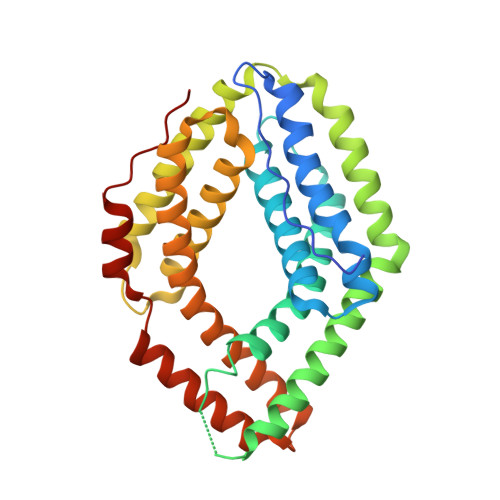
    Inorganic polyphosphates (polyPs) are linear polymers of orthophosphate units linked by phosphoanhydride bonds. Here, we report that bacterial, archaeal, and eukaryotic conserved histidine α-helical (CHAD) domains are specific polyP-binding modules. Crystal structures reveal that CHAD domains are formed by two four-helix bundles, giving rise to a central pore surrounded by conserved basic surface patches. Different CHAD domains bind polyPs with dissociation constants ranging from the nano- to mid-micromolar range, but not nucleic acids. A CHAD-polyP complex structure reveals the phosphate polymer binding across the central pore and along the two basic patches. Mutational analysis of CHAD-polyP interface residues validates the complex structure. The presence of a CHAD domain in the polyPase ygiF enhances its enzymatic activity. The only known CHAD protein from the plant Ricinus communis localizes to the nucleus/nucleolus when expressed in Arabidopsis and tobacco, suggesting that plants may harbor polyPs in these compartments. We propose that CHAD domains may be used to engineer the properties of polyP-metabolizing enzymes and to specifically localize polyP stores in eukaryotic cells and tissues.


  • Organizational Affiliation
    • Structural Plant Biology Laboratory, Department of Botany and Plant Biology, Faculty of Sciences, University of Geneva, Geneva, Switzerland.

Macromolecule Content 

  • Total Structure Weight: 33.34 kDa 
  • Atom Count: 2,269 
  • Modeled Residue Count: 289 
  • Deposited Residue Count: 303 
  • Unique protein chains: 1

Macromolecules

Find similar proteins by:|  3D Structure
Entity ID: 1
MoleculeChains  Sequence LengthOrganismDetailsImage
CHAD domain303Ricinus communisMutation(s): 0 
Gene Names: RCOM_0386350
UniProt
Find proteins for B9T8Q5 (Ricinus communis)
Explore B9T8Q5 
Go to UniProtKB:  B9T8Q5
Entity Groups
Sequence Clusters30% Identity50% Identity70% Identity90% Identity95% Identity100% Identity
UniProt GroupB9T8Q5
Sequence Annotations
Expand
Reference Sequence

Experimental Data & Validation

Experimental Data

  • Method: X-RAY DIFFRACTION
  • Resolution: 2.30 Å
  • R-Value Free:  0.242 (Depositor), 0.224 (DCC) 
  • R-Value Work:  0.232 (Depositor) 
  • R-Value Observed: 0.232 (Depositor) 
Space Group: P 61 2 2
Unit Cell:
Length ( Å )Angle ( ˚ )
a = 116.427α = 90
b = 116.427β = 90
c = 119.248γ = 120
Software Package:
Software NamePurpose
BUSTERrefinement
XDSdata reduction
XDSdata scaling
SHARPphasing

Structure Validation

View Full Validation Report


Entry History 

& Funding Information

Deposition Data


Funding OrganizationLocationGrant Number
European Research CouncilSwitzerland310856

Revision History  (Full details and data files)

  • Version 1.0: 2019-05-29
    Type: Initial release
  • Version 1.1: 2019-06-05
    Changes: Data collection, Database references
  • Version 1.2: 2024-05-15
    Changes: Data collection, Database references, Derived calculations