6QUN | pdb_00006qun

Crystal structure of AtGapC1 with the catalytic Cys149 irreversibly oxidized by H2O2 treatment


Experimental Data Snapshot

  • Method: X-RAY DIFFRACTION
  • Resolution: 3.00 Å
  • R-Value Free: 
    0.292 (Depositor), 0.296 (DCC) 
  • R-Value Work: 
    0.228 (Depositor), 0.238 (DCC) 
  • R-Value Observed: 
    0.231 (Depositor) 

Starting Model: experimental
View more details

wwPDB Validation 3D Report Full Report

Validation slider image for 6QUN

Ligand Structure Quality Assessment 


This is version 1.3 of the entry. See complete history

Literature

Glutathionylation primes soluble glyceraldehyde-3-phosphate dehydrogenase for late collapse into insoluble aggregates.

Zaffagnini, M.Marchand, C.H.Malferrari, M.Murail, S.Bonacchi, S.Genovese, D.Montalti, M.Venturoli, G.Falini, G.Baaden, M.Lemaire, S.D.Fermani, S.Trost, P.

(2019) Proc Natl Acad Sci U S A 116: 26057-26065

  • DOI: https://doi.org/10.1073/pnas.1914484116
  • Primary Citation Related Structures: 
    6QUN, 6QUQ

  • PubMed Abstract: 

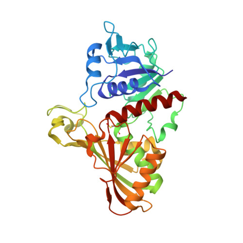
    Protein aggregation is a complex physiological process, primarily determined by stress-related factors revealing the hidden aggregation propensity of proteins that otherwise are fully soluble. Here we report a mechanism by which glycolytic glyceraldehyde-3-phosphate dehydrogenase of Arabidopsis thaliana (AtGAPC1) is primed to form insoluble aggregates by the glutathionylation of its catalytic cysteine (Cys149). Following a lag phase, glutathionylated AtGAPC1 initiates a self-aggregation process resulting in the formation of branched chains of globular particles made of partially misfolded and totally inactive proteins. GSH molecules within AtGAPC1 active sites are suggested to provide the initial destabilizing signal. The following removal of glutathione by the formation of an intramolecular disulfide bond between Cys149 and Cys153 reinforces the aggregation process. Physiological reductases, thioredoxins and glutaredoxins, could not dissolve AtGAPC1 aggregates but could efficiently contrast their growth. Besides acting as a protective mechanism against overoxidation, S-glutathionylation of AtGAPC1 triggers an unexpected aggregation pathway with completely different and still unexplored physiological implications.


  • Organizational Affiliation
    • Department of Pharmacy and Biotechnology, University of Bologna, 40126 Bologna, Italy; mirko.zaffagnini3@unibo.it paolo.trost@unibo.it.

Macromolecule Content 

  • Total Structure Weight: 75.1 kDa 
  • Atom Count: 5,296 
  • Modeled Residue Count: 668 
  • Deposited Residue Count: 668 
  • Unique protein chains: 1

Macromolecules

Find similar proteins by:|  3D Structure
Entity ID: 1
MoleculeChains  Sequence LengthOrganismDetailsImage
Glyceraldehyde-3-phosphate dehydrogenase GAPC1, cytosolicA [auth O],
B [auth R]
334Arabidopsis thalianaMutation(s): 0 
Gene Names: GAPC1GAPCGAPDHAt3g04120T6K12.26
EC: 1.2.1.12
UniProt
Find proteins for P25858 (Arabidopsis thaliana)
Explore P25858 
Go to UniProtKB:  P25858
Entity Groups
Sequence Clusters30% Identity50% Identity70% Identity90% Identity95% Identity100% Identity
UniProt GroupP25858
Sequence Annotations
Expand
Reference Sequence

Small Molecules

Ligands 2 Unique
IDChains Name / Formula / InChI Key2D Diagram3D Interactions
NAD

Query on NAD


Download:Ideal Coordinates CCD File
C [auth O],
G [auth R]
NICOTINAMIDE-ADENINE-DINUCLEOTIDE
C21 H27 N7 O14 P2
BAWFJGJZGIEFAR-NNYOXOHSSA-N
SO4

Query on SO4


Download:Ideal Coordinates CCD File
D [auth O]
E [auth O]
F [auth O]
H [auth R]
I [auth R]
D [auth O],
E [auth O],
F [auth O],
H [auth R],
I [auth R],
J [auth R],
K [auth R]
SULFATE ION
O4 S
QAOWNCQODCNURD-UHFFFAOYSA-L
Modified Residues  1 Unique
IDChains TypeFormula2D DiagramParent
CSD
Query on CSD
A [auth O],
B [auth R]
L-PEPTIDE LINKINGC3 H7 N O4 SCYS

Experimental Data & Validation

Experimental Data

  • Method: X-RAY DIFFRACTION
  • Resolution: 3.00 Å
  • R-Value Free:  0.292 (Depositor), 0.296 (DCC) 
  • R-Value Work:  0.228 (Depositor), 0.238 (DCC) 
  • R-Value Observed: 0.231 (Depositor) 
Space Group: P 61 2 2
Unit Cell:
Length ( Å )Angle ( ˚ )
a = 77.677α = 90
b = 77.677β = 90
c = 407.346γ = 120
Software Package:
Software NamePurpose
PHENIXrefinement
XDSdata reduction
SCALAdata scaling
MOLREPphasing

Structure Validation

View Full Validation Report


Ligand Structure Quality Assessment 


Entry History 

& Funding Information

Deposition Data


Funding OrganizationLocationGrant Number
ItalyFARB2012-University of Bologna

Revision History  (Full details and data files)

  • Version 1.0: 2019-12-04
    Type: Initial release
  • Version 1.1: 2019-12-25
    Changes: Database references
  • Version 1.2: 2024-01-24
    Changes: Data collection, Database references, Refinement description
  • Version 1.3: 2024-10-23
    Changes: Structure summary