6QUG | pdb_00006qug

GHK tagged MBP-Nup98(1-29)


Experimental Data Snapshot

  • Method: X-RAY DIFFRACTION
  • Resolution: 2.70 Å
  • R-Value Free: 
    0.226 (Depositor), 0.227 (DCC) 
  • R-Value Work: 
    0.207 (Depositor), 0.208 (DCC) 
  • R-Value Observed: 
    0.208 (Depositor) 

wwPDB Validation 3D Report Full Report

Validation slider image for 6QUG

This is version 3.2 of the entry. See complete history

Literature

The copper(II)-binding tripeptide GHK, a valuable crystallization and phasing tag for macromolecular crystallography.

Mehr, A.Henneberg, F.Chari, A.Gorlich, D.Huyton, T.

(2020) Acta Crystallogr D Struct Biol 76: 1222-1232

  • DOI: https://doi.org/10.1107/S2059798320013741
  • Primary Citation Related Structures: 
    6QUG, 6QUH, 6QUI, 6QUJ

  • PubMed Abstract: 

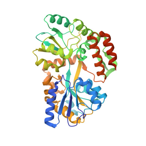
    The growth of diffraction-quality crystals and experimental phasing remain two of the main bottlenecks in protein crystallography. Here, the high-affinity copper(II)-binding tripeptide GHK was fused to the N-terminus of a GFP variant and an MBP-FG peptide fusion. The GHK tag promoted crystallization, with various residues (His, Asp, His/Pro) from symmetry molecules completing the copper(II) square-pyramidal coordination sphere. Rapid structure determination by copper SAD phasing could be achieved, even at a very low Bijvoet ratio or after significant radiation damage. When collecting highly redundant data at a wavelength close to the copper absorption edge, residual S-atom positions could also be located in log-likelihood-gradient maps and used to improve the phases. The GHK copper SAD method provides a convenient way of both crystallizing and phasing macromolecular structures, and will complement the current trend towards native sulfur SAD and MR-SAD phasing.


  • Organizational Affiliation
    • Department of Structural Dynamics, Max Planck Institute for Biophysical Chemistry, Göttingen, Germany.

Macromolecule Content 

  • Total Structure Weight: 525.75 kDa 
  • Atom Count: 34,893 
  • Modeled Residue Count: 4,474 
  • Deposited Residue Count: 4,824 
  • Unique protein chains: 1

Macromolecules

Find similar proteins by:|  3D Structure
Entity ID: 1
MoleculeChains  Sequence LengthOrganismDetailsImage
Maltodextrin-binding protein,Nucleoporin, putative
A, B, C, D, E
A, B, C, D, E, F, G, H, I, J, K, L
402Escherichia coliTetrahymena thermophila SB210
This entity is chimeric
Mutation(s): 0 
Gene Names: malENCTC8450_00456NCTC9775_03059TTHERM_00071070
UniProt
Find proteins for I7MHJ5 (Tetrahymena thermophila (strain SB210))
Explore I7MHJ5 
Go to UniProtKB:  I7MHJ5
Find proteins for P0AEX9 (Escherichia coli (strain K12))
Explore P0AEX9 
Go to UniProtKB:  P0AEX9
Entity Groups
Sequence Clusters30% Identity50% Identity70% Identity90% Identity95% Identity100% Identity
UniProt GroupsI7MHJ5P0AEX9
Sequence Annotations
Expand
Reference Sequence

Small Molecules

Ligands 3 Unique
IDChains Name / Formula / InChI Key2D Diagram3D Interactions
SO4

Query on SO4


Download:Ideal Coordinates CCD File
BA [auth C],
OA [auth K],
QA [auth L]
SULFATE ION
O4 S
QAOWNCQODCNURD-UHFFFAOYSA-L
PO4

Query on PO4


Download:Ideal Coordinates CCD File
DA [auth D],
EA [auth D],
KA [auth I],
MA [auth J]
PHOSPHATE ION
O4 P
NBIIXXVUZAFLBC-UHFFFAOYSA-K
CU

Query on CU


Download:Ideal Coordinates CCD File
AA [auth C]
CA [auth D]
FA [auth E]
GA [auth F]
HA [auth G]
AA [auth C],
CA [auth D],
FA [auth E],
GA [auth F],
HA [auth G],
IA [auth H],
JA [auth I],
LA [auth J],
NA [auth K],
PA [auth L],
Y [auth A],
Z [auth B]
COPPER (II) ION
Cu
JPVYNHNXODAKFH-UHFFFAOYSA-N

Experimental Data & Validation

Experimental Data

  • Method: X-RAY DIFFRACTION
  • Resolution: 2.70 Å
  • R-Value Free:  0.226 (Depositor), 0.227 (DCC) 
  • R-Value Work:  0.207 (Depositor), 0.208 (DCC) 
  • R-Value Observed: 0.208 (Depositor) 
Space Group: P 1 21 1
Unit Cell:
Length ( Å )Angle ( ˚ )
a = 82.91α = 90
b = 286.78β = 92.01
c = 110.78γ = 90
Software Package:
Software NamePurpose
REFMACrefinement
XDSdata reduction
XSCALEdata scaling
CRANK2phasing

Structure Validation

View Full Validation Report


Entry History 

Deposition Data

Revision History  (Full details and data files)

  • Version 1.0: 2020-05-27
    Type: Initial release
  • Version 2.0: 2020-07-29
    Type: Remediation
    Reason: Carbohydrate remediation
    Changes: Advisory, Atomic model, Data collection, Derived calculations, Structure summary
  • Version 3.0: 2020-12-09
    Type: Coordinate replacement
    Reason: Atomic clashes
    Changes: Advisory, Atomic model, Data collection, Database references, Derived calculations, Non-polymer description, Refinement description, Structure summary
  • Version 3.1: 2020-12-16
    Changes: Database references
  • Version 3.2: 2024-05-15
    Changes: Data collection, Database references, Refinement description