6QNZ | pdb_00006qnz

Crystal structure of the site-specific DNA nickase N.BspD6I E418A Mutant


Experimental Data Snapshot

  • Method: X-RAY DIFFRACTION
  • Resolution: 2.45 Å
  • R-Value Free: 
    0.243 (Depositor), 0.247 (DCC) 
  • R-Value Work: 
    0.231 (Depositor), 0.234 (DCC) 
  • R-Value Observed: 
    0.232 (Depositor) 

Starting Model: experimental
View more details

wwPDB Validation 3D Report Full Report

Validation slider image for 6QNZ

This is version 1.3 of the entry. See complete history

Literature

The key role of E418 carboxyl group in the formation of Nt.BspD6I nickase active site: Structural and functional properties of Nt.BspD6I E418A mutant.

Artyukh, R.I.Kachalova, G.S.Yunusova, A.K.Fatkhullin, B.F.Atanasov, B.P.Perevyazova, T.A.Popov, A.N.Gabdulkhakov, A.G.Zheleznaya, L.A.

(2020) J Struct Biol 210: 107508-107508

  • DOI: https://doi.org/10.1016/j.jsb.2020.107508
  • Primary Citation Related Structures: 
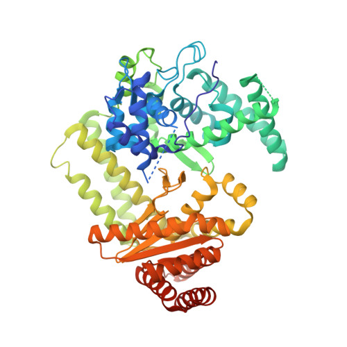
    6QNZ

  • PubMed Abstract: 

    The mutated nickase Nt.BspD6I E418A has been obtained by site-directed mutagenesis. The purified protein has been crystallized, and its spatial structure has been determined at 2.45 Å resolution. An analysis of the crystal structures of the wild-type and mutated nickase have shown that the elimination of a carboxyl group due to the E418A mutation initiates marked conformational changes in both the N-terminal recognition domain and the C-terminal catalytic domain of nickase and insignificantly affects its linker domain. This is supported by changes in the functional properties of mutated nickase: an increase in the oligomerization capacity in the presence of a substrate, a reduction in the capacity to bind a substrate, and complete loss of catalytic activity.


  • Organizational Affiliation
    • Institute of Theoretical and Experimental Biophysics, Russian Academy of Sciences, Pushchino, Moscow Region 142290, Russia. Electronic address: rimmaartyukh@gmail.com.

Macromolecule Content 

  • Total Structure Weight: 142.29 kDa 
  • Atom Count: 9,515 
  • Modeled Residue Count: 1,142 
  • Deposited Residue Count: 1,208 
  • Unique protein chains: 1

Macromolecules

Find similar proteins by:|  3D Structure
Entity ID: 1
MoleculeChains  Sequence LengthOrganismDetailsImage
Heterodimeric restriction endonuclease R.BspD6I large subunit
A, B
604Bacillus sp. D6Mutation(s): 1 
Gene Names: bspD6R1
UniProt
Find proteins for A3FEV7 (Bacillus sp. D6)
Explore A3FEV7 
Go to UniProtKB:  A3FEV7
Entity Groups
Sequence Clusters30% Identity50% Identity70% Identity90% Identity95% Identity100% Identity
UniProt GroupA3FEV7
Sequence Annotations
Expand
Reference Sequence

Experimental Data & Validation

Experimental Data

  • Method: X-RAY DIFFRACTION
  • Resolution: 2.45 Å
  • R-Value Free:  0.243 (Depositor), 0.247 (DCC) 
  • R-Value Work:  0.231 (Depositor), 0.234 (DCC) 
  • R-Value Observed: 0.232 (Depositor) 
Space Group: P 1 21 1
Unit Cell:
Length ( Å )Angle ( ˚ )
a = 76.43α = 90
b = 92.25β = 105.61
c = 113.8γ = 90
Software Package:
Software NamePurpose
PHENIXrefinement
XDSdata reduction
XSCALEdata scaling
PHASERphasing

Structure Validation

View Full Validation Report


Entry History 

Revision History  (Full details and data files)

  • Version 1.0: 2020-03-04
    Type: Initial release
  • Version 1.1: 2020-04-29
    Changes: Database references
  • Version 1.2: 2020-05-20
    Changes: Database references
  • Version 1.3: 2024-01-24
    Changes: Data collection, Database references, Refinement description