6QCB | pdb_00006qcb

Crystal structure of human cathepsin D in complex with macrocyclic inhibitor 9


Experimental Data Snapshot

  • Method: X-RAY DIFFRACTION
  • Resolution: 1.55 Å
  • R-Value Free: 
    0.190 (Depositor), 0.195 (DCC) 
  • R-Value Work: 
    0.166 (Depositor), 0.172 (DCC) 
  • R-Value Observed: 
    0.167 (Depositor) 

Starting Model: experimental
View more details

wwPDB Validation 3D Report Full Report

Validation slider image for 6QCB

Ligand Structure Quality Assessment 


This is version 2.2 of the entry. See complete history

Literature

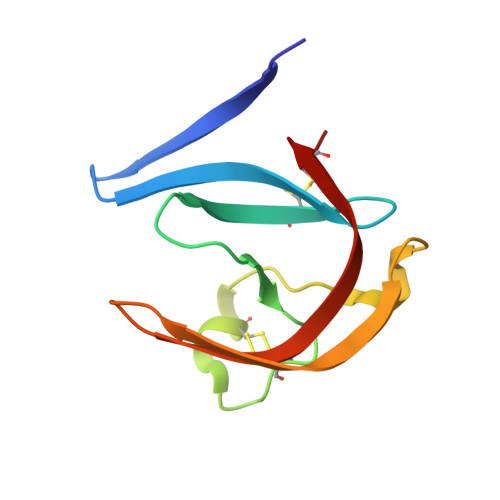
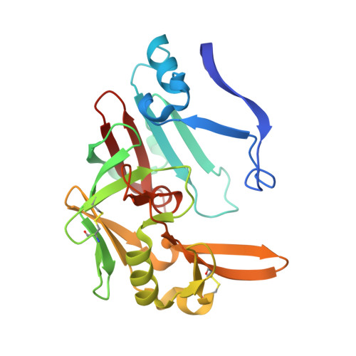
Biomimetic Macrocyclic Inhibitors of Human Cathepsin D: Structure-Activity Relationship and Binding Mode Analysis.

Houstecka, R.Hadzima, M.Fanfrlik, J.Brynda, J.Pallova, L.Hanova, I.Mertlikova-Kaiserova, H.Lepsik, M.Horn, M.Smrcina, M.Majer, P.Mares, M.

(2020) J Med Chem 63: 1576-1596

  • DOI: https://doi.org/10.1021/acs.jmedchem.9b01351
  • Primary Citation Related Structures: 
    6QBG, 6QBH, 6QCB

  • PubMed Abstract: 

    Human cathepsin D (CatD), a pepsin-family aspartic protease, plays an important role in tumor progression and metastasis. Here, we report the development of biomimetic inhibitors of CatD as novel tools for regulation of this therapeutic target. We designed a macrocyclic scaffold to mimic the spatial conformation of the minimal pseudo-dipeptide binding motif of pepstatin A, a microbial oligopeptide inhibitor, in the CatD active site. A library of more than 30 macrocyclic peptidomimetic inhibitors was employed for scaffold optimization, mapping of subsite interactions, and profiling of inhibitor selectivity. Furthermore, we solved high-resolution crystal structures of three macrocyclic inhibitors with low nanomolar or subnanomolar potency in complex with CatD and determined their binding mode using quantum chemical calculations. The study provides a new structural template and functional profile that can be exploited for design of potential chemotherapeutics that specifically inhibit CatD and related aspartic proteases.


  • Organizational Affiliation
    • Institute of Organic Chemistry and Biochemistry , Czech Academy of Sciences , Flemingovo nám. 2 , 16610 Praha 6 , Czech Republic.

Macromolecule Content 

  • Total Structure Weight: 38.91 kDa 
  • Atom Count: 3,152 
  • Modeled Residue Count: 338 
  • Deposited Residue Count: 338 
  • Unique protein chains: 2

Macromolecules

Find similar proteins by:|  3D Structure
Entity ID: 1
MoleculeChains  Sequence LengthOrganismDetailsImage
Cathepsin D97Homo sapiensMutation(s): 0 
Gene Names: CTSDCPSD
EC: 3.4.23.5
UniProt & NIH Common Fund Data Resources
Find proteins for P07339 (Homo sapiens)
Explore P07339 
Go to UniProtKB:  P07339
PHAROS:  P07339
GTEx:  ENSG00000117984 
Entity Groups
Sequence Clusters30% Identity50% Identity70% Identity90% Identity95% Identity100% Identity
UniProt GroupP07339
Glycosylation
Glycosylation Sites: 1Go to GlyGen: P07339-1
Sequence Annotations
Expand
Reference Sequence
Find similar proteins by:|  3D Structure
Entity ID: 2
MoleculeChains  Sequence LengthOrganismDetailsImage
Cathepsin D241Homo sapiensMutation(s): 0 
Gene Names: CTSDCPSD
EC: 3.4.23.5
UniProt & NIH Common Fund Data Resources
Find proteins for P07339 (Homo sapiens)
Explore P07339 
Go to UniProtKB:  P07339
PHAROS:  P07339
GTEx:  ENSG00000117984 
Entity Groups
Sequence Clusters30% Identity50% Identity70% Identity90% Identity95% Identity100% Identity
UniProt GroupP07339
Glycosylation
Glycosylation Sites: 1Go to GlyGen: P07339-1
Sequence Annotations
Expand
Reference Sequence

Oligosaccharides

Help  
Entity ID: 3
MoleculeChains Length2D Diagram GlycosylationD Interactions
2-acetamido-2-deoxy-beta-D-glucopyranose-(1-4)-2-acetamido-2-deoxy-beta-D-glucopyranose
C
2N-Glycosylation
Glycosylation Resources
GlyTouCan: G42666HT
GlyCosmos: G42666HT
GlyGen: G42666HT
Entity ID: 4
MoleculeChains Length2D Diagram GlycosylationD Interactions
beta-D-mannopyranose-(1-4)-2-acetamido-2-deoxy-beta-D-glucopyranose-(1-4)-2-acetamido-2-deoxy-beta-D-glucopyranose
D
3N-Glycosylation
Glycosylation Resources
GlyTouCan: G15407YE
GlyCosmos: G15407YE
GlyGen: G15407YE

Small Molecules

Ligands 4 Unique
IDChains Name / Formula / InChI Key2D Diagram3D Interactions
HWE

Query on HWE


Download:Ideal Coordinates CCD File
F [auth A](3~{S},7~{S},8~{S})-7-oxidanyl-8-(phenylmethyl)-3-propan-2-yl-1,4,9-triazacyclohenicosane-2,5,10-trione
C28 H45 N3 O4
FTRRUFDJLIEMLS-DPZBCOQUSA-N
SO4

Query on SO4


Download:Ideal Coordinates CCD File
H [auth B],
I [auth B]
SULFATE ION
O4 S
QAOWNCQODCNURD-UHFFFAOYSA-L
PO3

Query on PO3


Download:Ideal Coordinates CCD File
E [auth A]PHOSPHITE ION
O3 P
AQSJGOWTSHOLKH-UHFFFAOYSA-N
DMS

Query on DMS


Download:Ideal Coordinates CCD File
G [auth A]DIMETHYL SULFOXIDE
C2 H6 O S
IAZDPXIOMUYVGZ-UHFFFAOYSA-N
Binding Affinity Annotations 
IDSourceBinding Affinity
HWE BindingDB:  6QCB IC50: 5.8 (nM) from 1 assay(s)

Experimental Data & Validation

Experimental Data

  • Method: X-RAY DIFFRACTION
  • Resolution: 1.55 Å
  • R-Value Free:  0.190 (Depositor), 0.195 (DCC) 
  • R-Value Work:  0.166 (Depositor), 0.172 (DCC) 
  • R-Value Observed: 0.167 (Depositor) 
Space Group: C 2 2 21
Unit Cell:
Length ( Å )Angle ( ˚ )
a = 80.346α = 90
b = 94.257β = 90
c = 117.675γ = 90
Software Package:
Software NamePurpose
REFMACrefinement
XDSdata reduction
XSCALEdata scaling
MOLREPphasing

Structure Validation

View Full Validation Report


Ligand Structure Quality Assessment 


Entry History 

& Funding Information

Deposition Data


Funding OrganizationLocationGrant Number
Czech RepublicRVO 61388963
European Regional Development FundCzech RepublicCZ.02.1.01/0.0/0.0/16_019/0000729
Czech Republic802218

Revision History  (Full details and data files)

  • Version 1.0: 2020-01-29
    Type: Initial release
  • Version 1.1: 2020-02-19
    Changes: Data collection, Database references
  • Version 1.2: 2020-03-11
    Changes: Database references
  • Version 2.0: 2020-07-29
    Type: Remediation
    Reason: Carbohydrate remediation
    Changes: Atomic model, Data collection, Derived calculations, Structure summary
  • Version 2.1: 2024-01-24
    Changes: Data collection, Database references, Refinement description, Structure summary
  • Version 2.2: 2024-11-13
    Changes: Structure summary