6OPC | pdb_00006opc

Cdc48 Hexamer in a complex with substrate and Shp1(Ubx Domain)


Experimental Data Snapshot

  • Method: ELECTRON MICROSCOPY
  • Resolution: 3.70 Å
  • Aggregation State: PARTICLE 
  • Reconstruction Method: SINGLE PARTICLE 

wwPDB Validation 3D Report Full Report

Validation slider image for 6OPC

This is version 1.3 of the entry. See complete history

Literature

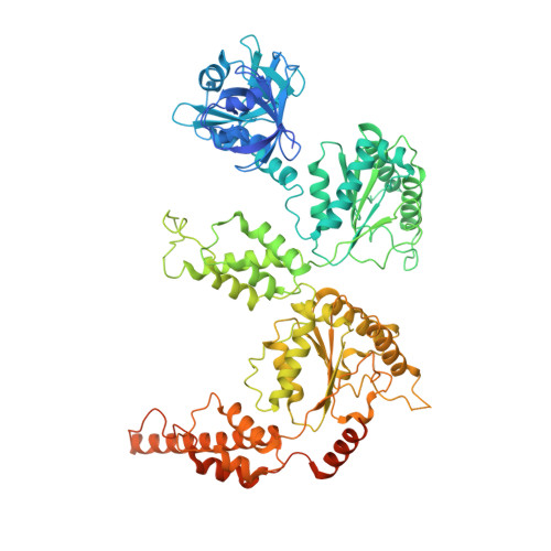
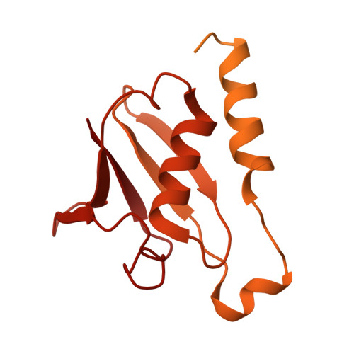
Structure of the Cdc48 segregase in the act of unfolding an authentic substrate.

Cooney, I.Han, H.Stewart, M.G.Carson, R.H.Hansen, D.T.Iwasa, J.H.Price, J.C.Hill, C.P.Shen, P.S.

(2019) Science 365: 502-505

  • DOI: https://doi.org/10.1126/science.aax0486
  • Primary Citation Related Structures: 
    6OMB, 6OPC

  • PubMed Abstract: 

    The cellular machine Cdc48 functions in multiple biological pathways by segregating its protein substrates from a variety of stable environments such as organelles or multi-subunit complexes. Despite extensive studies, the mechanism of Cdc48 has remained obscure, and its reported structures are inconsistent with models of substrate translocation proposed for other AAA+ ATPases (adenosine triphosphatases). Here, we report a 3.7-angstrom-resolution structure of Cdc48 in complex with an adaptor protein and a native substrate. Cdc48 engages substrate by adopting a helical configuration of substrate-binding residues that extends through the central pore of both of the ATPase rings. These findings indicate a unified hand-over-hand mechanism of protein translocation by Cdc48 and other AAA+ ATPases.


  • Organizational Affiliation
    • Department of Biochemistry, 15 N. Medical Drive East, University of Utah, Salt Lake City, UT 84112, USA.

Macromolecule Content 

  • Total Structure Weight: 606.57 kDa 
  • Atom Count: 30,015 
  • Modeled Residue Count: 4,477 
  • Deposited Residue Count: 5,455 
  • Unique protein chains: 3

Macromolecules

Find similar proteins by:|  3D Structure
Entity ID: 1
MoleculeChains  Sequence LengthOrganismDetailsImage
Cell division control protein 48
A, B, C, D, E
A, B, C, D, E, F
835Saccharomyces cerevisiaeMutation(s): 0 
EC: 3.6.4.6
UniProt
Find proteins for P25694 (Saccharomyces cerevisiae (strain ATCC 204508 / S288c))
Explore P25694 
Go to UniProtKB:  P25694
Entity Groups
Sequence Clusters30% Identity50% Identity70% Identity90% Identity95% Identity100% Identity
UniProt GroupP25694
Sequence Annotations
Expand
Reference Sequence
Find similar proteins by:  Sequence   |   3D Structure  
Entity ID: 2
MoleculeChains  Sequence LengthOrganismDetailsImage
Substrate bound to the central pore of the Cdc48 hexamer22Saccharomyces cerevisiaeMutation(s): 0 
Find similar proteins by:|  3D Structure
Entity ID: 3
MoleculeChains  Sequence LengthOrganismDetailsImage
UBX domain-containing protein 1H [auth Z]423Saccharomyces cerevisiaeMutation(s): 0 
UniProt
Find proteins for P34223 (Saccharomyces cerevisiae (strain ATCC 204508 / S288c))
Explore P34223 
Go to UniProtKB:  P34223
Entity Groups
Sequence Clusters30% Identity50% Identity70% Identity90% Identity95% Identity100% Identity
UniProt GroupP34223
Sequence Annotations
Expand
Reference Sequence

Small Molecules

Ligands 3 Unique
IDChains Name / Formula / InChI Key2D Diagram3D Interactions
ADP

Query on ADP


Download:Ideal Coordinates CCD File
AA [auth D]
DA [auth D]
GA [auth E]
HA [auth E]
I [auth A]
AA [auth D],
DA [auth D],
GA [auth E],
HA [auth E],
I [auth A],
L [auth A],
O [auth B],
Q [auth B],
U [auth C],
X [auth C]
ADENOSINE-5'-DIPHOSPHATE
C10 H15 N5 O10 P2
XTWYTFMLZFPYCI-KQYNXXCUSA-N
BEF

Query on BEF


Download:Ideal Coordinates CCD File
BA [auth D]
EA [auth D]
J [auth A]
M [auth A]
R [auth B]
BA [auth D],
EA [auth D],
J [auth A],
M [auth A],
R [auth B],
T [auth C],
V [auth C],
Y [auth C]
BERYLLIUM TRIFLUORIDE ION
Be F3
OGIAHMCCNXDTIE-UHFFFAOYSA-K
MG

Query on MG


Download:Ideal Coordinates CCD File
CA [auth D]
FA [auth D]
K [auth A]
N [auth A]
P [auth B]
CA [auth D],
FA [auth D],
K [auth A],
N [auth A],
P [auth B],
S [auth B],
W [auth C],
Z [auth C]
MAGNESIUM ION
Mg
JLVVSXFLKOJNIY-UHFFFAOYSA-N

Experimental Data & Validation

Experimental Data

  • Method: ELECTRON MICROSCOPY
  • Resolution: 3.70 Å
  • Aggregation State: PARTICLE 
  • Reconstruction Method: SINGLE PARTICLE 

Structure Validation

View Full Validation Report


Entry History 

& Funding Information

Deposition Data


Funding OrganizationLocationGrant Number
National Institutes of Health/National Human Genome Research Institute (NIH/NHGRI)United StatesP50GM082545

Revision History  (Full details and data files)

  • Version 1.0: 2019-07-10
    Type: Initial release
  • Version 1.1: 2019-08-14
    Changes: Data collection, Database references
  • Version 1.2: 2019-12-18
    Changes: Author supporting evidence
  • Version 1.3: 2024-03-20
    Changes: Data collection, Database references, Derived calculations