6ON3 | pdb_00006on3

A substrate bound structure of L-DOPA dioxygenase from Streptomyces sclerotialus


Experimental Data Snapshot

  • Method: X-RAY DIFFRACTION
  • Resolution: 2.31 Å
  • R-Value Free: 
    0.294 (Depositor), 0.297 (DCC) 
  • R-Value Work: 
    0.235 (Depositor), 0.236 (DCC) 

wwPDB Validation 3D Report Full Report

Validation slider image for 6ON3

This is version 1.3 of the entry. See complete history

Literature

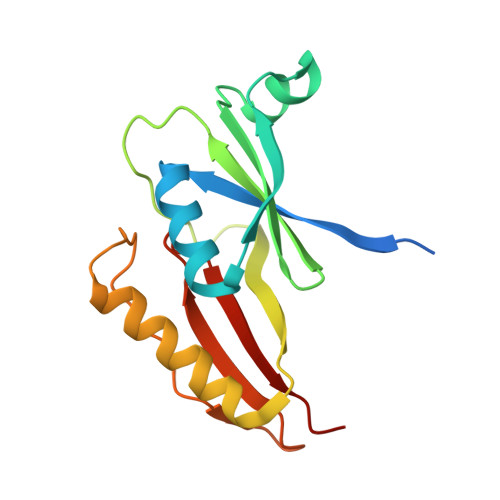
Crystal Structures of L-DOPA Dioxygenase fromStreptomyces sclerotialus.

Wang, Y.Shin, I.Fu, Y.Colabroy, K.L.Liu, A.

(2019) Biochemistry 58: 5339-5350

  • DOI: https://doi.org/10.1021/acs.biochem.9b00396
  • Primary Citation Related Structures: 
    6ON1, 6ON3

  • PubMed Abstract: 

    Extradiol dioxygenases are essential biocatalysts for breaking down catechols. The vicinal oxygen chelate (VOC) superfamily contains a large number of extradiol dioxygenases, most of which are found as part of catabolic pathways degrading a variety of natural and human-made aromatic rings. The l-3,4-dihydroxyphenylalanine (L-DOPA) extradiol dioxygenases compose a multitude of pathways that produce various antibacterial or antitumor natural products. The structural features of these dioxygenases are anticipated to be distinct from those of other VOC extradiol dioxygenases. Herein, we identified a new L-DOPA dioxygenase from the thermophilic bacterium Streptomyces sclerotialus (SsDDO) through a sequence and genome context analysis. The activity of SsDDO was kinetically characterized with L-DOPA using an ultraviolet-visible spectrophotometer and an oxygen electrode. The optimal temperature of the assay was 55 °C, at which the K m and k cat of SsDDO were 110 ± 10 μM and 2.0 ± 0.1 s -1 , respectively. We determined the de novo crystal structures of SsDDO in the ligand-free form and as a substrate-bound complex, refined to 1.99 and 2.31 Å resolution, respectively. These structures reveal that SsDDO possesses a form IV arrangement of βαβββ modules, the first characterization of this assembly from among the VOC/type I extradiol dioxygenase protein family. Electron paramagnetic resonance spectra of Fe-NO adducts for the resting and substrate-bound enzyme were obtained. This work contributes to our understanding of a growing class of topologically distinct VOC dioxygenases, and the obtained structural features will improve our understanding of the extradiol cleavage reaction within the VOC superfamily.


  • Organizational Affiliation
    • Department of Chemistry , University of Texas at San Antonio , San Antonio , Texas 78249 , United States.

Macromolecule Content 

  • Total Structure Weight: 115.3 kDa 
  • Atom Count: 7,678 
  • Modeled Residue Count: 879 
  • Deposited Residue Count: 990 
  • Unique protein chains: 1

Macromolecules

Find similar proteins by:|  3D Structure
Entity ID: 1
MoleculeChains  Sequence LengthOrganismDetailsImage
L-DOPA extradiol dioxygenase
A, B, C, D, E
A, B, C, D, E, F
165Streptomyces sclerotialusMutation(s): 0 
UniProt
Find proteins for A0A5H1ZR51 (Streptomyces sclerotialus)
Explore A0A5H1ZR51 
Go to UniProtKB:  A0A5H1ZR51
Entity Groups
Sequence Clusters30% Identity50% Identity70% Identity90% Identity95% Identity100% Identity
UniProt GroupA0A5H1ZR51
Sequence Annotations
Expand
Reference Sequence

Small Molecules

Ligands 3 Unique
IDChains Name / Formula / InChI Key2D Diagram3D Interactions
DAH

Query on DAH


Download:Ideal Coordinates CCD File
I [auth A]
N [auth B]
O [auth C]
R [auth D]
T [auth E]
I [auth A],
N [auth B],
O [auth C],
R [auth D],
T [auth E],
W [auth F]
3,4-DIHYDROXYPHENYLALANINE
C9 H11 N O4
WTDRDQBEARUVNC-LURJTMIESA-N
TRS

Query on TRS


Download:Ideal Coordinates CCD File
G [auth A],
H [auth A],
L [auth B],
M [auth B],
V [auth F]
2-AMINO-2-HYDROXYMETHYL-PROPANE-1,3-DIOL
C4 H12 N O3
LENZDBCJOHFCAS-UHFFFAOYSA-O
FE2

Query on FE2


Download:Ideal Coordinates CCD File
J [auth A]
K [auth B]
P [auth C]
Q [auth D]
S [auth E]
J [auth A],
K [auth B],
P [auth C],
Q [auth D],
S [auth E],
U [auth F]
FE (II) ION
Fe
CWYNVVGOOAEACU-UHFFFAOYSA-N

Experimental Data & Validation

Experimental Data

  • Method: X-RAY DIFFRACTION
  • Resolution: 2.31 Å
  • R-Value Free:  0.294 (Depositor), 0.297 (DCC) 
  • R-Value Work:  0.235 (Depositor), 0.236 (DCC) 
Space Group: P 1 21 1
Unit Cell:
Length ( Å )Angle ( ˚ )
a = 99.83α = 90
b = 40.72β = 105.11
c = 128.43γ = 90
Software Package:
Software NamePurpose
PHENIXrefinement
DENZOdata reduction
SCALEPACKdata scaling
PDB_EXTRACTdata extraction
PHENIXphasing

Structure Validation

View Full Validation Report


Entry History 

& Funding Information

Deposition Data


Funding OrganizationLocationGrant Number
National Science Foundation (NSF, United States)United StatesCHE-1808637
National Science Foundation (NSF, United States)United StatesCHE-1708237
National Institutes of Health/National Institute of General Medical Sciences (NIH/NIGMS)United StatesR01GM108988

Revision History  (Full details and data files)

  • Version 1.0: 2019-06-26
    Type: Initial release
  • Version 1.1: 2019-11-27
    Changes: Author supporting evidence
  • Version 1.2: 2020-01-08
    Changes: Database references
  • Version 1.3: 2024-03-13
    Changes: Data collection, Database references, Derived calculations