6O6E | pdb_00006o6e

Crystal structure of PltF trapped with PltL using a proline adenosine vinylsulfonamide inhibitor


Experimental Data Snapshot

  • Method: X-RAY DIFFRACTION
  • Resolution: 2.14 Å
  • R-Value Free: 
    0.260 (Depositor), 0.262 (DCC) 
  • R-Value Work: 
    0.214 (Depositor), 0.218 (DCC) 
  • R-Value Observed: 
    0.216 (Depositor) 

Starting Model: experimental
View more details

wwPDB Validation 3D Report Full Report

Validation slider image for 6O6E

Ligand Structure Quality Assessment 


This is version 3.2 of the entry. See complete history

Literature

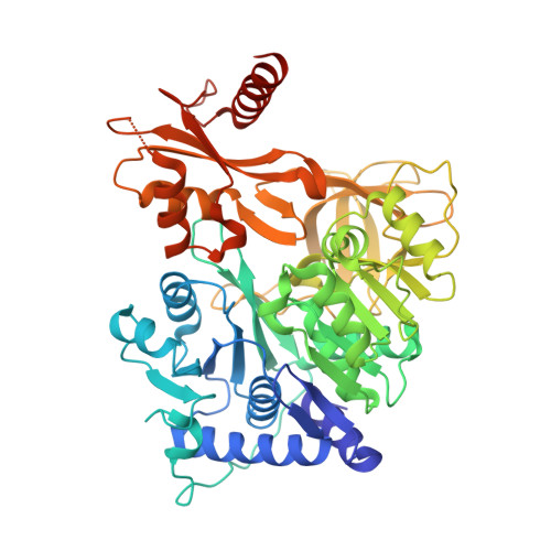
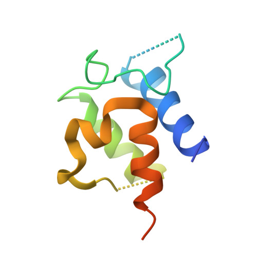
Dynamic visualization of type II peptidyl carrier protein recognition in pyoluteorin biosynthesis.

Corpuz, J.C.Podust, L.M.Davis, T.D.Jaremko, M.J.Burkart, M.D.

(2020) RSC Chem Biol 1: 8-12

  • DOI: https://doi.org/10.1039/c9cb00015a
  • Primary Citation Related Structures: 
    6O6E

  • PubMed Abstract: 

    Using a covalent chemical probe and X-ray crystallography coupled to nuclear magnetic resonance data, we elucidated the dynamic molecular basis of protein recognition between the carrier protein and adenylation domain in pyoluteorin biosynthesis. These findings reveal a unique binding mode, which contrasts previously solved carrier protein and partner protein interfaces.


  • Organizational Affiliation
    • Department of Chemistry and Biochemistry, University of California-San Diego, 9500 Gilman Drive, La Jolla, CA 92093-0358, USA.

Macromolecule Content 

  • Total Structure Weight: 69.23 kDa 
  • Atom Count: 4,552 
  • Modeled Residue Count: 578 
  • Deposited Residue Count: 609 
  • Unique protein chains: 2

Macromolecules

Find similar proteins by:|  3D Structure
Entity ID: 1
MoleculeChains  Sequence LengthOrganismDetailsImage
L-proline--[L-prolyl-carrier protein] ligaseA [auth B]513Pseudomonas protegens Pf-5Mutation(s): 0 
Gene Names: pltFPFL_2792
EC: 6.2.1.53
UniProt
Find proteins for Q4KCY5 (Pseudomonas fluorescens (strain ATCC BAA-477 / NRRL B-23932 / Pf-5))
Explore Q4KCY5 
Go to UniProtKB:  Q4KCY5
Entity Groups
Sequence Clusters30% Identity50% Identity70% Identity90% Identity95% Identity100% Identity
UniProt GroupQ4KCY5
Sequence Annotations
Expand
Reference Sequence
Find similar proteins by:|  3D Structure
Entity ID: 2
MoleculeChains  Sequence LengthOrganismDetailsImage
Peptidyl carrier protein PltLB [auth E]96Pseudomonas protegens Pf-5Mutation(s): 0 
Gene Names: pltLPFL_2786
UniProt
Find proteins for Q4KCZ1 (Pseudomonas fluorescens (strain ATCC BAA-477 / NRRL B-23932 / Pf-5))
Explore Q4KCZ1 
Go to UniProtKB:  Q4KCZ1
Entity Groups
Sequence Clusters30% Identity50% Identity70% Identity90% Identity95% Identity100% Identity
UniProt GroupQ4KCZ1
Sequence Annotations
Expand
Reference Sequence

Small Molecules

Ligands 4 Unique
IDChains Name / Formula / InChI Key2D Diagram3D Interactions
I5M
(Subject of Investigation/LOI)

Query on I5M


Download:Ideal Coordinates CCD File
S [auth E]5'-deoxy-5'-({(2S)-2-({2-[(N-{(2R)-4-[(dioxo-lambda~5~-phosphanyl)oxy]-2-hydroxy-3,3-dimethylbutanoyl}-beta-alanyl)amino]ethyl}sulfanyl)-2-[(2S)-pyrrolidin-2-yl]ethanesulfonyl}amino)adenosine
C27 H44 N9 O11 P S2
UUZQPBPCNSKBOR-LCZLERPASA-N
TRS

Query on TRS


Download:Ideal Coordinates CCD File
P [auth B]2-AMINO-2-HYDROXYMETHYL-PROPANE-1,3-DIOL
C4 H12 N O3
LENZDBCJOHFCAS-UHFFFAOYSA-O
MLA

Query on MLA


Download:Ideal Coordinates CCD File
Q [auth B],
R [auth B]
MALONIC ACID
C3 H4 O4
OFOBLEOULBTSOW-UHFFFAOYSA-N
FMT

Query on FMT


Download:Ideal Coordinates CCD File
C [auth B]
D [auth B]
E [auth B]
F [auth B]
G [auth B]
C [auth B],
D [auth B],
E [auth B],
F [auth B],
G [auth B],
H [auth B],
I [auth B],
J [auth B],
K [auth B],
L [auth B],
M [auth B],
N [auth B],
O [auth B]
FORMIC ACID
C H2 O2
BDAGIHXWWSANSR-UHFFFAOYSA-N

Experimental Data & Validation

Experimental Data

  • Method: X-RAY DIFFRACTION
  • Resolution: 2.14 Å
  • R-Value Free:  0.260 (Depositor), 0.262 (DCC) 
  • R-Value Work:  0.214 (Depositor), 0.218 (DCC) 
  • R-Value Observed: 0.216 (Depositor) 
Space Group: P 32 2 1
Unit Cell:
Length ( Å )Angle ( ˚ )
a = 170.55α = 90
b = 170.55β = 90
c = 64.9γ = 120
Software Package:
Software NamePurpose
REFMACrefinement
XDSdata reduction
XDSdata scaling
MOLREPphasing

Structure Validation

View Full Validation Report


Ligand Structure Quality Assessment 


Entry History 

& Funding Information

Deposition Data


Funding OrganizationLocationGrant Number
National Institutes of Health/National Human Genome Research Institute (NIH/NHGRI)United StatesT32 GM008326
National Institutes of Health/National Human Genome Research Institute (NIH/NHGRI)United StatesR01 GM095970

Revision History  (Full details and data files)

  • Version 1.0: 2020-04-08
    Type: Initial release
  • Version 1.1: 2020-06-17
    Changes: Database references
  • Version 2.0: 2020-07-22
    Type: Coordinate replacement
    Reason: Sequence discrepancy
    Changes: Advisory, Atomic model, Author supporting evidence, Data collection, Database references, Derived calculations, Polymer sequence, Refinement description, Source and taxonomy, Structure summary
  • Version 2.1: 2020-11-11
    Changes: Database references
  • Version 2.2: 2020-12-23
    Changes: Database references
  • Version 3.0: 2022-07-20
    Type: Coordinate replacement
    Reason: Ligand geometry
    Changes: Advisory, Atomic model, Author supporting evidence, Data collection, Database references, Derived calculations, Non-polymer description, Refinement description, Source and taxonomy, Structure summary
  • Version 3.1: 2023-10-11
    Changes: Data collection, Refinement description
  • Version 3.2: 2024-10-23
    Changes: Structure summary