6O3L | pdb_00006o3l

Crystal structure of the Fab fragment of the human HIV-1 neutralizing antibody PGZL1.H4K3 in complex with its MPER peptide epitope (region 671-683 of HIV-1 gp41).


Experimental Data Snapshot

  • Method: X-RAY DIFFRACTION
  • Resolution: 1.98 Å
  • R-Value Free: 
    0.211 (Depositor), 0.210 (DCC) 
  • R-Value Work: 
    0.170 (Depositor), 0.172 (DCC) 
  • R-Value Observed: 
    0.172 (Depositor) 

Starting Model: experimental
View more details

wwPDB Validation 3D Report Full Report

Validation slider image for 6O3L

This is version 1.3 of the entry. See complete history

Literature

An MPER antibody neutralizes HIV-1 using germline features shared among donors.

Zhang, L.Irimia, A.He, L.Landais, E.Rantalainen, K.Leaman, D.P.Vollbrecht, T.Stano, A.Sands, D.I.Kim, A.S.Poignard, P.Burton, D.R.Murrell, B.Ward, A.B.Zhu, J.Wilson, I.A.Zwick, M.B.

(2019) Nat Commun 10: 5389-5389

  • DOI: https://doi.org/10.1038/s41467-019-12973-1
  • Primary Citation Related Structures: 
    6O3D, 6O3G, 6O3J, 6O3K, 6O3L, 6O3U, 6O41, 6O42

  • PubMed Abstract: 

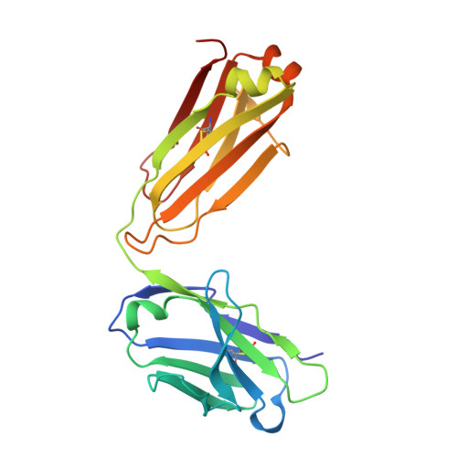
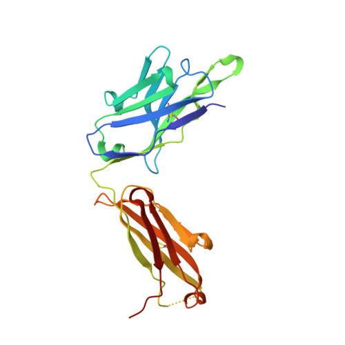
    The membrane-proximal external region (MPER) of HIV-1 envelope glycoprotein (Env) can be targeted by neutralizing antibodies of exceptional breadth. MPER antibodies usually have long, hydrophobic CDRH3s, lack activity as inferred germline precursors, are often from the minor IgG3 subclass, and some are polyreactive, such as 4E10. Here we describe an MPER broadly neutralizing antibody from the major IgG1 subclass, PGZL1, which shares germline V/D-region genes with 4E10, has a shorter CDRH3, and is less polyreactive. A recombinant sublineage variant pan-neutralizes a 130-isolate panel at 1.4 μg/ml (IC 50 ). Notably, a germline revertant with mature CDR3s neutralizes 12% of viruses and still binds MPER after DJ reversion. Crystal structures of lipid-bound PGZL1 variants and cryo-EM reconstruction of an Env-PGZL1 complex reveal how these antibodies recognize MPER and viral membrane. Discovery of common genetic and structural elements among MPER antibodies from different patients suggests that such antibodies could be elicited using carefully designed immunogens.


  • Organizational Affiliation
    • Department of Immunology and Microbiology, The Scripps Research Institute, La Jolla, California, 92037, USA.

Macromolecule Content 

  • Total Structure Weight: 100.4 kDa 
  • Atom Count: 7,628 
  • Modeled Residue Count: 905 
  • Deposited Residue Count: 916 
  • Unique protein chains: 3

Macromolecules

Find similar proteins by:|  3D Structure
Entity ID: 1
MoleculeChains  Sequence LengthOrganismDetailsImage
PGZL1.H4K3 light chainA [auth L],
C [auth B]
215Homo sapiensMutation(s): 0 
Find similar proteins by:|  3D Structure
Entity ID: 2
MoleculeChains  Sequence LengthOrganismDetailsImage
PGZL1.H4K3 heavy chainB [auth H],
D [auth A]
227Homo sapiensMutation(s): 0 
Find similar proteins by:  Sequence   |   3D Structure  
Entity ID: 3
MoleculeChains  Sequence LengthOrganismDetailsImage
MPER peptide, region 671-683 of HIV-1 gp41E [auth D],
F [auth E]
16Human immunodeficiency virus 1Mutation(s): 0 
UniProt
Find proteins for P04578 (Human immunodeficiency virus type 1 group M subtype B (isolate HXB2))
Explore P04578 
Go to UniProtKB:  P04578
Entity Groups
Sequence Clusters30% Identity50% Identity70% Identity90% Identity95% Identity100% Identity
UniProt GroupP04578
Sequence Annotations
Expand
Reference Sequence

Small Molecules

Ligands 2 Unique
IDChains Name / Formula / InChI Key2D Diagram3D Interactions
PO4

Query on PO4


Download:Ideal Coordinates CCD File
I [auth H]
J [auth H]
K [auth H]
M [auth B]
N [auth A]
I [auth H],
J [auth H],
K [auth H],
M [auth B],
N [auth A],
O [auth A]
PHOSPHATE ION
O4 P
NBIIXXVUZAFLBC-UHFFFAOYSA-K
GOL

Query on GOL


Download:Ideal Coordinates CCD File
G [auth L],
H [auth L],
L [auth H],
P [auth A],
Q [auth E]
GLYCEROL
C3 H8 O3
PEDCQBHIVMGVHV-UHFFFAOYSA-N

Experimental Data & Validation

Experimental Data

  • Method: X-RAY DIFFRACTION
  • Resolution: 1.98 Å
  • R-Value Free:  0.211 (Depositor), 0.210 (DCC) 
  • R-Value Work:  0.170 (Depositor), 0.172 (DCC) 
  • R-Value Observed: 0.172 (Depositor) 
Space Group: P 1
Unit Cell:
Length ( Å )Angle ( ˚ )
a = 55.342α = 87.94
b = 58.029β = 76.09
c = 98.725γ = 72.52
Software Package:
Software NamePurpose
PHENIXrefinement
HKL-2000data reduction
HKL-2000data scaling
PHASERphasing

Structure Validation

View Full Validation Report


Entry History 

& Funding Information

Deposition Data


Funding OrganizationLocationGrant Number
National Institutes of Health/National Institute Of Allergy and Infectious Diseases (NIH/NIAID)United StatesUM1 AI100663

Revision History  (Full details and data files)

  • Version 1.0: 2019-12-04
    Type: Initial release
  • Version 1.1: 2019-12-11
    Changes: Database references
  • Version 1.2: 2023-10-11
    Changes: Data collection, Database references, Refinement description
  • Version 1.3: 2024-11-06
    Changes: Structure summary