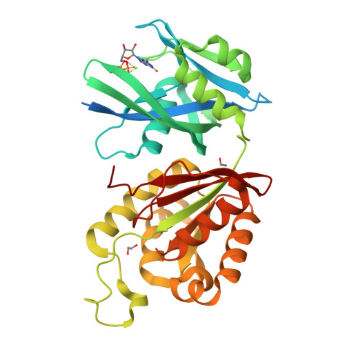
RCSB PDB - 6M69: Crystal structure of Mycobacterium smegmatis MutT1 in complex with GMPPCP (GDP)

 6M69

Crystal structure of Mycobacterium smegmatis MutT1 in complex with GMPPCP (GDP)


Experimental Data Snapshot

  • Method: X-RAY DIFFRACTION
  • Resolution: 1.50 Å
  • R-Value Free: 0.223 
  • R-Value Work: 0.188 
  • R-Value Observed: 0.190 

Starting Model: experimental
View more details

wwPDB Validation   3D Report Full Report


Ligand Structure Quality Assessment 

Created with Raphaël 2.3.0Worse 01 BetterLigand structure goodness of fit to experimental dataBest fitted GDPClick on this verticalbar to view detailsBest fitted POPClick on this verticalbar to view details

This is version 1.2 of the entry. See complete history


Literature

Plasticity, ligand conformation and enzyme action of Mycobacterium smegmatis MutT1.

Raj, P.Karthik, S.Arif, S.M.Varshney, U.Vijayan, M.

(2020) Acta Crystallogr D Struct Biol 76: 982-992

  • DOI: https://doi.org/10.1107/S2059798320010992
  • Primary Citation of Related Structures:  
    6M65, 6M69, 6M6Y, 6M72

  • PubMed Abstract: 

    Mycobacterium smegmatis MutT1 (MsMutT1) is a sanitation enzyme made up of an N-terminal Nudix hydrolase domain and a C-terminal domain resembling a histidine phosphatase. It has been established that the action of MutT1 on 8-oxo-dGTP, 8-oxo-GTP and diadenosine polyphosphates is modulated by intermolecular interactions. In order to further explore this and to elucidate the structural basis of its differential action on 8-oxo-NTPs and unsubstituted NTPs, the crystal structures of complexes of MsMutT1 with 8-oxo-dGTP, GMPPNP and GMPPCP have been determined. Replacement soaking was used in order to ensure that the complexes were isomorphous to one another. Analysis of the structural data led to the elucidation of a relationship between the arrangements of molecules observed in the crystals, molecular plasticity and the action of the enzyme on nucleotides. The dominant mode of arrangement involving a head-to-tail sequence predominantly leads to the generation of NDPs. The other mode of packing arrangement appears to preferentially generate NMPs. This work also provides interesting insights into the dependence of enzyme action on the conformation of the ligand. The possibility of modulating the enzyme action through differences in intermolecular interactions and ligand conformations makes MsMutT1 a versatile enzyme.


  • Organizational Affiliation

    Molecular Biophysics Unit, Indian Institute of Science, Bengaluru 560 012, India.


Macromolecules
Find similar proteins by:  (by identity cutoff)  |  3D Structure
Entity ID: 1
MoleculeChains Sequence LengthOrganismDetailsImage
Hydrolase, NUDIX family protein342Mycolicibacterium smegmatis MC2 155Mutation(s): 0 
Gene Names: MSMEG_2390
EC: 3.6.1.61 (UniProt), 3.6.1.69 (UniProt), 3.6.1.58 (UniProt)
UniProt
Find proteins for A0QUZ2 (Mycolicibacterium smegmatis (strain ATCC 700084 / mc(2)155))
Explore A0QUZ2 
Go to UniProtKB:  A0QUZ2
Entity Groups  
Sequence Clusters30% Identity50% Identity70% Identity90% Identity95% Identity100% Identity
UniProt GroupA0QUZ2
Sequence Annotations
Expand
  • Reference Sequence
Small Molecules
Ligands 4 Unique
IDChains Name / Formula / InChI Key2D Diagram3D Interactions
GDP (Subject of Investigation/LOI)
Query on GDP

Download Ideal Coordinates CCD File 
B [auth A]GUANOSINE-5'-DIPHOSPHATE
C10 H15 N5 O11 P2
QGWNDRXFNXRZMB-UUOKFMHZSA-N
POP (Subject of Investigation/LOI)
Query on POP

Download Ideal Coordinates CCD File 
C [auth A]PYROPHOSPHATE 2-
H2 O7 P2
XPPKVPWEQAFLFU-UHFFFAOYSA-L
EDO
Query on EDO

Download Ideal Coordinates CCD File 
D [auth A],
E [auth A]
1,2-ETHANEDIOL
C2 H6 O2
LYCAIKOWRPUZTN-UHFFFAOYSA-N
MG (Subject of Investigation/LOI)
Query on MG

Download Ideal Coordinates CCD File 
F [auth A]MAGNESIUM ION
Mg
JLVVSXFLKOJNIY-UHFFFAOYSA-N
Experimental Data & Validation

Experimental Data

  • Method: X-RAY DIFFRACTION
  • Resolution: 1.50 Å
  • R-Value Free: 0.223 
  • R-Value Work: 0.188 
  • R-Value Observed: 0.190 
  • Space Group: P 21 21 2
Unit Cell:
Length ( Å )Angle ( ˚ )
a = 192.65α = 90
b = 37.08β = 90
c = 44.8γ = 90
Software Package:
Software NamePurpose
MOSFLMdata reduction
SCALAdata scaling
PHASERphasing
REFMACrefinement
PDB_EXTRACTdata extraction

Structure Validation

View Full Validation Report


Ligand Structure Quality Assessment 

Created with Raphaël 2.3.0Worse 01 BetterLigand structure goodness of fit to experimental dataBest fitted GDPClick on this verticalbar to view detailsBest fitted POPClick on this verticalbar to view details

Entry History & Funding Information

Deposition Data


Funding OrganizationLocationGrant Number
Department of Biotechnology (DBT, India)IndiaBT/PR20529/BRB/10/1525/2016

Revision History  (Full details and data files)

  • Version 1.0: 2020-10-14
    Type: Initial release
  • Version 1.1: 2020-10-28
    Changes: Database references
  • Version 1.2: 2023-11-29
    Changes: Data collection, Database references, Refinement description