6IUF | pdb_00006iuf

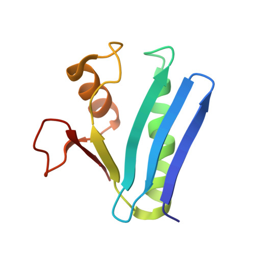
Crystal structure of Anti-CRISPR protein AcrVA5


Experimental Data Snapshot

  • Method: X-RAY DIFFRACTION
  • Resolution: 2.05 Å
  • R-Value Free: 
    0.186 (Depositor), 0.188 (DCC) 
  • R-Value Work: 
    0.152 (Depositor) 
  • R-Value Observed: 
    0.154 (Depositor) 

wwPDB Validation 3D Report Full Report

Validation slider image for 6IUF

Ligand Structure Quality Assessment 


This is version 1.2 of the entry. See complete history. 

Literature

An anti-CRISPR protein disables type V Cas12a by acetylation.

Dong, L., Guan, X., Li, N., Zhang, F., Zhu, Y., Ren, K., Yu, L., Zhou, F., Han, Z., Gao, N., Huang, Z.

(2019) Nat Struct Mol Biol 26: 308-314

  • DOI: https://doi.org/10.1038/s41594-019-0206-1
  • Primary Citation Related Structures: 
    6IUF, 6IV6

  • PubMed Abstract: 

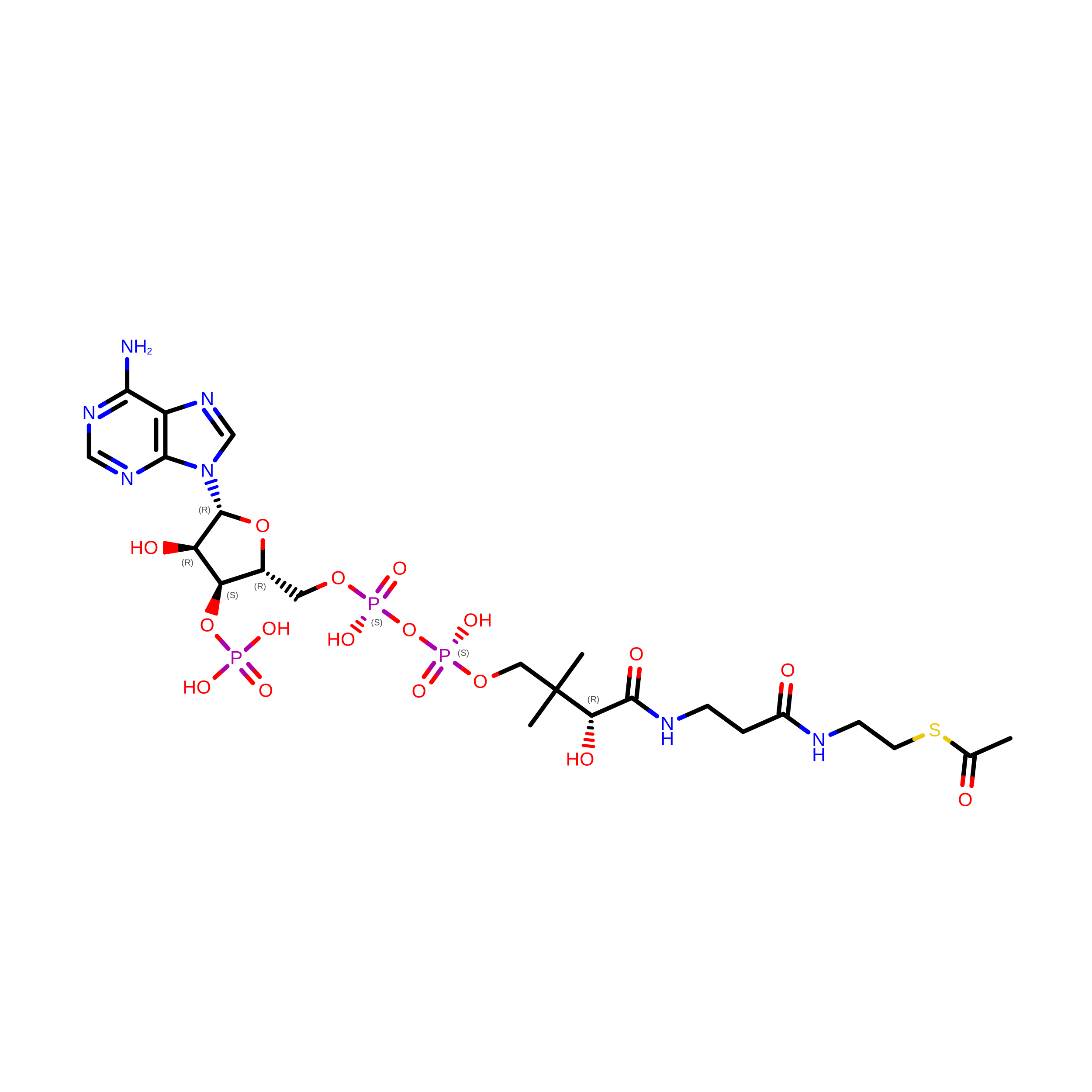
    Phages use anti-CRISPR proteins to deactivate the CRISPR-Cas system. The mechanisms for the inhibition of type I and type II systems by anti-CRISPRs have been elucidated. However, it has remained unknown how the type V CRISPR-Cas12a (Cpf1) system is inhibited by anti-CRISPRs. Here we identify the anti-CRISPR protein AcrVA5 and report the mechanisms by which it inhibits CRISPR-Cas12a. Our structural and biochemical data show that AcrVA5 functions as an acetyltransferase to modify Moraxella bovoculi (Mb) Cas12a at Lys635, a residue that is required for recognition of the protospacer-adjacent motif. The AcrVA5-mediated modification of MbCas12a results in complete loss of double-stranded DNA (dsDNA)-cleavage activity. In contrast, the Lys635Arg mutation renders MbCas12a completely insensitive to inhibition by AcrVA5. A cryo-EM structure of the AcrVA5-acetylated MbCas12a reveals that Lys635 acetylation provides sufficient steric hindrance to prevent dsDNA substrates from binding to the Cas protein. Our study reveals an unprecedented mechanism of CRISPR-Cas inhibition and suggests an evolutionary arms race between phages and bacteria.


  • Organizational Affiliation: 
    • HIT Center for Life Sciences, School of Life Science and Technology, Harbin Institute of Technology, Harbin, China.

Macromolecule Content 

  • Total Structure Weight: 23.57 kDa 
  • Atom Count: 1,769 
  • Modeled Residue Count: 191 
  • Deposited Residue Count: 192 
  • Unique protein chains: 1

Macromolecules

Find similar proteins by:|  3D Structure
Entity ID: 1
MoleculeChains  Sequence LengthOrganismDetailsImage
protein-a
A, B
96Moraxella bovoculiMutation(s): 0 
Gene Names: AAX07_09540
Entity Groups
Sequence Clusters30% Identity50% Identity70% Identity90% Identity95% Identity100% Identity
Sequence Annotations
Expand
Reference Sequence

Experimental Data & Validation

Experimental Data

  • Method: X-RAY DIFFRACTION
  • Resolution: 2.05 Å
  • R-Value Free:  0.186 (Depositor), 0.188 (DCC) 
  • R-Value Work:  0.152 (Depositor) 
  • R-Value Observed: 0.154 (Depositor) 
Space Group: P 41 3 2
Unit Cell:
Length ( Å )Angle ( ˚ )
a = 143.422α = 90
b = 143.422β = 90
c = 143.422γ = 90
Software Package:
Software NamePurpose
PHENIXrefinement
HKL-2000data reduction
SCALAdata scaling
PHASERphasing

Structure Validation

View Full Validation Report


Ligand Structure Quality Assessment 


Entry History 

& Funding Information

Deposition Data


Funding OrganizationLocationGrant Number
National Natural Science Foundation of ChinaChina31825008
National Natural Science Foundation of ChinaChina31422014

Revision History  (Full details and data files)

  • Version 1.0: 2019-04-10
    Type: Initial release
  • Version 1.1: 2019-04-17
    Changes: Data collection, Database references
  • Version 1.2: 2024-03-27
    Changes: Data collection, Database references