6I8B | pdb_00006i8b

Crystal structure of Spindlin1 in complex with the inhibitor VinSpinIn


Experimental Data Snapshot

  • Method: X-RAY DIFFRACTION
  • Resolution: 1.76 Å
  • R-Value Free: 
    0.254 (Depositor), 0.259 (DCC) 
  • R-Value Work: 
    0.233 (Depositor), 0.237 (DCC) 
  • R-Value Observed: 
    0.234 (Depositor) 

Starting Model: experimental
View more details

wwPDB Validation 3D Report Full Report

Validation slider image for 6I8B

Ligand Structure Quality Assessment 


This is version 1.2 of the entry. See complete history

Literature

A Chemical Probe for Tudor Domain Protein Spindlin1 to Investigate Chromatin Function.

Fagan, V.Johansson, C.Gileadi, C.Monteiro, O.Dunford, J.E.Nibhani, R.Philpott, M.Malzahn, J.Wells, G.Faram, R.Cribbs, A.P.Halidi, N.Li, F.Chau, I.Greschik, H.Velupillai, S.Allali-Hassani, A.Bennett, J.Christott, T.Giroud, C.Lewis, A.M.Huber, K.V.M.Athanasou, N.Bountra, C.Jung, M.Schule, R.Vedadi, M.Arrowsmith, C.Xiong, Y.Jin, J.Fedorov, O.Farnie, G.Brennan, P.E.Oppermann, U.

(2019) J Med Chem 62: 9008-9025

  • DOI: https://doi.org/10.1021/acs.jmedchem.9b00562
  • Primary Citation Related Structures: 
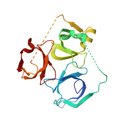
    6I8B, 6I8L, 6I8Y

  • PubMed Abstract: 

    Modifications of histone tails, including lysine/arginine methylation, provide the basis of a "chromatin or histone code". Proteins that contain "reader" domains can bind to these modifications and form specific effector complexes, which ultimately mediate chromatin function. The spindlin1 (SPIN1) protein contains three Tudor methyllysine/arginine reader domains and was identified as a putative oncogene and transcriptional coactivator. Here we report a SPIN1 chemical probe inhibitor with low nanomolar in vitro activity, exquisite selectivity on a panel of methyl reader and writer proteins, and with submicromolar cellular activity. X-ray crystallography showed that this Tudor domain chemical probe simultaneously engages Tudor domains 1 and 2 via a bidentate binding mode. Small molecule inhibition and siRNA knockdown of SPIN1, as well as chemoproteomic studies, identified genes which are transcriptionally regulated by SPIN1 in squamous cell carcinoma and suggest that SPIN1 may have a role in cancer related inflammation and/or cancer metastasis.


  • Organizational Affiliation
    • Structural Genomics Consortium, Nuffield Department of Medicine , University of Oxford , OX3 7DQ Oxford , U.K.

Macromolecule Content 

  • Total Structure Weight: 53.11 kDa 
  • Atom Count: 3,293 
  • Modeled Residue Count: 387 
  • Deposited Residue Count: 444 
  • Unique protein chains: 1

Macromolecules

Find similar proteins by:|  3D Structure
Entity ID: 1
MoleculeChains  Sequence LengthOrganismDetailsImage
Spindlin-1A [auth B],
B [auth E]
222Homo sapiensMutation(s): 0 
Gene Names: SPIN1OCRSPIN
UniProt & NIH Common Fund Data Resources
Find proteins for Q9Y657 (Homo sapiens)
Explore Q9Y657 
Go to UniProtKB:  Q9Y657
PHAROS:  Q9Y657
GTEx:  ENSG00000106723 
Entity Groups
Sequence Clusters30% Identity50% Identity70% Identity90% Identity95% Identity100% Identity
UniProt GroupQ9Y657
Sequence Annotations
Expand
Reference Sequence

Small Molecules

Ligands 4 Unique
IDChains Name / Formula / InChI Key2D Diagram3D Interactions
H7T

Query on H7T


Download:Ideal Coordinates CCD File
C [auth B],
J [auth E]
2-[4-[2-[[2-[3-[2-azanyl-5-(cyclopropylmethoxy)-3,3-dimethyl-indol-6-yl]oxypropyl]-1,3-dihydroisoindol-5-yl]oxy]ethyl]-1,2,3-triazol-1-yl]-1-[4-(2-pyrrolidin-1-ylethyl)piperidin-1-yl]ethanone
C42 H58 N8 O4
XPEJZXWPKDAYFX-UHFFFAOYSA-N
PO4

Query on PO4


Download:Ideal Coordinates CCD File
I [auth E]PHOSPHATE ION
O4 P
NBIIXXVUZAFLBC-UHFFFAOYSA-K
DMS

Query on DMS


Download:Ideal Coordinates CCD File
D [auth B],
E [auth B],
F [auth B],
G [auth B],
K [auth E]
DIMETHYL SULFOXIDE
C2 H6 O S
IAZDPXIOMUYVGZ-UHFFFAOYSA-N
GLY

Query on GLY


Download:Ideal Coordinates CCD File
H [auth B],
L [auth E]
GLYCINE
C2 H5 N O2
DHMQDGOQFOQNFH-UHFFFAOYSA-N
Binding Affinity Annotations 
IDSourceBinding Affinity
H7T BindingDB:  6I8B Kd: min: 9.9, max: 131 (nM) from 4 assay(s)
IC50: 33 (nM) from 1 assay(s)
EC50: 270 (nM) from 1 assay(s)

Experimental Data & Validation

Experimental Data

  • Method: X-RAY DIFFRACTION
  • Resolution: 1.76 Å
  • R-Value Free:  0.254 (Depositor), 0.259 (DCC) 
  • R-Value Work:  0.233 (Depositor), 0.237 (DCC) 
  • R-Value Observed: 0.234 (Depositor) 
Space Group: P 21 21 21
Unit Cell:
Length ( Å )Angle ( ˚ )
a = 111.875α = 90
b = 118.583β = 90
c = 43.808γ = 90
Software Package:
Software NamePurpose
XDSdata reduction
Aimlessdata scaling
PHASERphasing
PHENIXrefinement
PDB_EXTRACTdata extraction

Structure Validation

View Full Validation Report


Ligand Structure Quality Assessment 


Entry History 

Deposition Data

Revision History  (Full details and data files)

  • Version 1.0: 2018-12-05
    Type: Initial release
  • Version 1.1: 2020-01-15
    Changes: Database references
  • Version 1.2: 2024-01-24
    Changes: Data collection, Database references, Refinement description