6H2U | pdb_00006h2u

Crystal structure of human METTL5-TRMT112 complex, the 18S rRNA m6A1832 methyltransferase at 1.6A resolution


Experimental Data Snapshot

  • Method: X-RAY DIFFRACTION
  • Resolution: 1.60 Å
  • R-Value Free: 
    0.215 (Depositor), 0.214 (DCC) 
  • R-Value Work: 
    0.186 (Depositor), 0.188 (DCC) 
  • R-Value Observed: 
    0.187 (Depositor) 

wwPDB Validation 3D Report Full Report

Validation slider image for 6H2U

Ligand Structure Quality Assessment 


This is version 1.2 of the entry. See complete history

Literature

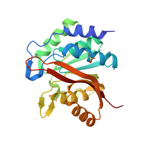
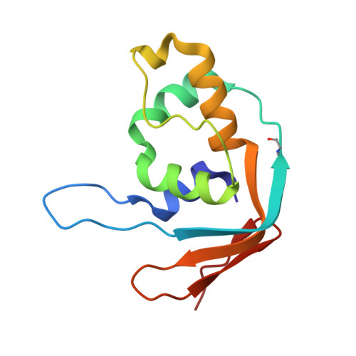
The human 18S rRNA m6A methyltransferase METTL5 is stabilized by TRMT112.

van Tran, N.Ernst, F.G.M.Hawley, B.R.Zorbas, C.Ulryck, N.Hackert, P.Bohnsack, K.E.Bohnsack, M.T.Jaffrey, S.R.Graille, M.Lafontaine, D.L.J.

(2019) Nucleic Acids Res 47: 7719-7733

  • DOI: https://doi.org/10.1093/nar/gkz619
  • Primary Citation Related Structures: 
    6H2U, 6H2V

  • PubMed Abstract: 

    N6-methyladenosine (m6A) has recently been found abundantly on messenger RNA and shown to regulate most steps of mRNA metabolism. Several important m6A methyltransferases have been described functionally and structurally, but the enzymes responsible for installing one m6A residue on each subunit of human ribosomes at functionally important sites have eluded identification for over 30 years. Here, we identify METTL5 as the enzyme responsible for 18S rRNA m6A modification and confirm ZCCHC4 as the 28S rRNA modification enzyme. We show that METTL5 must form a heterodimeric complex with TRMT112, a known methyltransferase activator, to gain metabolic stability in cells. We provide the first atomic resolution structure of METTL5-TRMT112, supporting that its RNA-binding mode differs distinctly from that of other m6A RNA methyltransferases. On the basis of similarities with a DNA methyltransferase, we propose that METTL5-TRMT112 acts by extruding the adenosine to be modified from a double-stranded nucleic acid.


  • Organizational Affiliation
    • BIOC, CNRS, Ecole polytechnique, Institut Polytechnique de Paris, F-91128 Palaiseau, France.

Macromolecule Content 

  • Total Structure Weight: 38.94 kDa 
  • Atom Count: 3,009 
  • Modeled Residue Count: 321 
  • Deposited Residue Count: 334 
  • Unique protein chains: 2

Macromolecules

Find similar proteins by:|  3D Structure
Entity ID: 1
MoleculeChains  Sequence LengthOrganismDetailsImage
Methyltransferase-like protein 5215Homo sapiensMutation(s): 0 
Gene Names: METTL5DC3HSPC133
EC: 2.1.1
UniProt & NIH Common Fund Data Resources
Find proteins for Q9NRN9 (Homo sapiens)
Explore Q9NRN9 
Go to UniProtKB:  Q9NRN9
PHAROS:  Q9NRN9
GTEx:  ENSG00000138382 
Entity Groups
Sequence Clusters30% Identity50% Identity70% Identity90% Identity95% Identity100% Identity
UniProt GroupQ9NRN9
Sequence Annotations
Expand
Reference Sequence
Find similar proteins by:|  3D Structure
Entity ID: 2
MoleculeChains  Sequence LengthOrganismDetailsImage
Multifunctional methyltransferase subunit TRM112-like protein118Homo sapiensMutation(s): 0 
Gene Names: TRMT112AD-001HSPC152HSPC170
UniProt & NIH Common Fund Data Resources
Find proteins for Q9UI30 (Homo sapiens)
Explore Q9UI30 
Go to UniProtKB:  Q9UI30
PHAROS:  Q9UI30
GTEx:  ENSG00000173113 
Entity Groups
Sequence Clusters30% Identity50% Identity70% Identity90% Identity95% Identity100% Identity
UniProt GroupQ9UI30
Sequence Annotations
Expand
Reference Sequence

Small Molecules

Ligands 3 Unique
IDChains Name / Formula / InChI Key2D Diagram3D Interactions
SAM

Query on SAM


Download:Ideal Coordinates CCD File
C [auth A]S-ADENOSYLMETHIONINE
C15 H22 N6 O5 S
MEFKEPWMEQBLKI-FCKMPRQPSA-N
SO4

Query on SO4


Download:Ideal Coordinates CCD File
G [auth A],
H [auth A]
SULFATE ION
O4 S
QAOWNCQODCNURD-UHFFFAOYSA-L
EDO

Query on EDO


Download:Ideal Coordinates CCD File
D [auth A],
E [auth A],
F [auth A],
I [auth B],
J [auth B]
1,2-ETHANEDIOL
C2 H6 O2
LYCAIKOWRPUZTN-UHFFFAOYSA-N
Modified Residues  2 Unique
IDChains TypeFormula2D DiagramParent
CSD
Query on CSD
A
L-PEPTIDE LINKINGC3 H7 N O4 SCYS
CSO
Query on CSO
A
L-PEPTIDE LINKINGC3 H7 N O3 SCYS

Experimental Data & Validation

Experimental Data

  • Method: X-RAY DIFFRACTION
  • Resolution: 1.60 Å
  • R-Value Free:  0.215 (Depositor), 0.214 (DCC) 
  • R-Value Work:  0.186 (Depositor), 0.188 (DCC) 
  • R-Value Observed: 0.187 (Depositor) 
Space Group: P 21 21 21
Unit Cell:
Length ( Å )Angle ( ˚ )
a = 56.887α = 90
b = 70.634β = 90
c = 84.776γ = 90
Software Package:
Software NamePurpose
XDSdata reduction
Aimlessdata scaling
PHASERphasing
PHENIXrefinement
PDB_EXTRACTdata extraction

Structure Validation

View Full Validation Report


Ligand Structure Quality Assessment 


Entry History 

& Funding Information

Deposition Data


Funding OrganizationLocationGrant Number
French National Research AgencyFranceANR-14-CE09-0016
French National Research AgencyFranceANR-16-CE11-0003

Revision History  (Full details and data files)

  • Version 1.0: 2019-07-31
    Type: Initial release
  • Version 1.1: 2019-09-18
    Changes: Data collection, Database references
  • Version 1.2: 2024-11-06
    Changes: Data collection, Database references, Structure summary