6FR9 | pdb_00006fr9

F11 T-Cell Receptor Recognising PKYVKQNTLKLAT Peptide Presented by HLA-DR*0101


Experimental Data Snapshot

  • Method: X-RAY DIFFRACTION
  • Resolution: 1.62 Å
  • R-Value Free: 
    0.211 (Depositor), 0.223 (DCC) 
  • R-Value Work: 
    0.179 (Depositor), 0.189 (DCC) 
  • R-Value Observed: 
    0.180 (Depositor) 

Starting Model: experimental
View more details

wwPDB Validation 3D Report Full Report

Validation slider image for 6FR9

This is version 1.3 of the entry. See complete history

Literature

In Silicoand Structural Analyses Demonstrate That Intrinsic Protein Motions Guide T Cell Receptor Complementarity Determining Region Loop Flexibility.

Holland, C.J.MacLachlan, B.J.Bianchi, V.Hesketh, S.J.Morgan, R.Vickery, O.Bulek, A.M.Fuller, A.Godkin, A.Sewell, A.K.Rizkallah, P.J.Wells, S.Cole, D.K.

(2018) Front Immunol 9: 674-674

  • DOI: https://doi.org/10.3389/fimmu.2018.00674
  • Primary Citation Related Structures: 
    6EH4, 6EH5, 6EH6, 6EH7, 6EH8, 6EH9, 6FR3, 6FR4, 6FR5, 6FR6, 6FR7, 6FR8, 6FR9, 6FRA, 6FRB, 6FRC, 6FUM, 6FUN, 6FUO, 6FUP, 6FUQ, 6FUR

  • PubMed Abstract: 

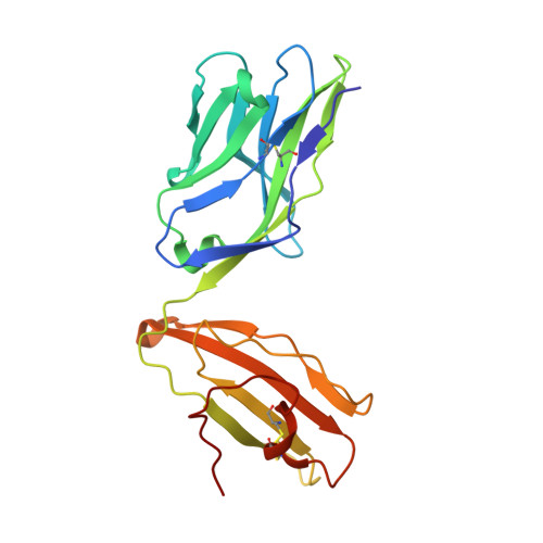
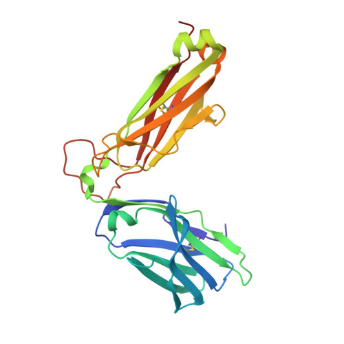
    T-cell immunity is controlled by T cell receptor (TCR) binding to peptide major histocompatibility complexes (pMHCs). The nature of the interaction between these two proteins has been the subject of many investigations because of its central role in immunity against pathogens, cancer, in autoimmunity, and during organ transplant rejection. Crystal structures comparing unbound and pMHC-bound TCRs have revealed flexibility at the interaction interface, particularly from the perspective of the TCR. However, crystal structures represent only a snapshot of protein conformation that could be influenced through biologically irrelevant crystal lattice contacts and other factors. Here, we solved the structures of three unbound TCRs from multiple crystals. Superposition of identical TCR structures from different crystals revealed some conformation differences of up to 5 Å in individual complementarity determining region (CDR) loops that are similar to those that have previously been attributed to antigen engagement. We then used a combination of rigidity analysis and simulations of protein motion to reveal the theoretical potential of TCR CDR loop flexibility in unbound state. These simulations of protein motion support the notion that crystal structures may only offer an artifactual indication of TCR flexibility, influenced by crystallization conditions and crystal packing that is inconsistent with the theoretical potential of intrinsic TCR motions.


  • Organizational Affiliation
    • Division of Infection and Immunity and Systems Immunity Research Institute, Cardiff University School of Medicine, Cardiff, United Kingdom.

Macromolecule Content 

  • Total Structure Weight: 50.33 kDa 
  • Atom Count: 4,062 
  • Modeled Residue Count: 442 
  • Deposited Residue Count: 442 
  • Unique protein chains: 2

Macromolecules

Find similar proteins by:|  3D Structure
Entity ID: 1
MoleculeChains  Sequence LengthOrganismDetailsImage
T-Cell Receptor F11 alpha chain202Homo sapiensMutation(s): 0 
Find similar proteins by:|  3D Structure
Entity ID: 2
MoleculeChains  Sequence LengthOrganismDetailsImage
T-Cell Receptor F11 beta chain240Homo sapiensMutation(s): 0 

Small Molecules

Ligands 3 Unique
IDChains Name / Formula / InChI Key2D Diagram3D Interactions
SO4

Query on SO4


Download:Ideal Coordinates CCD File
L [auth B]SULFATE ION
O4 S
QAOWNCQODCNURD-UHFFFAOYSA-L
GOL

Query on GOL


Download:Ideal Coordinates CCD File
K [auth B]GLYCEROL
C3 H8 O3
PEDCQBHIVMGVHV-UHFFFAOYSA-N
EDO

Query on EDO


Download:Ideal Coordinates CCD File
C [auth A]
D [auth A]
E [auth A]
F [auth A]
G [auth A]
C [auth A],
D [auth A],
E [auth A],
F [auth A],
G [auth A],
H [auth B],
I [auth B],
J [auth B]
1,2-ETHANEDIOL
C2 H6 O2
LYCAIKOWRPUZTN-UHFFFAOYSA-N

Experimental Data & Validation

Experimental Data

  • Method: X-RAY DIFFRACTION
  • Resolution: 1.62 Å
  • R-Value Free:  0.211 (Depositor), 0.223 (DCC) 
  • R-Value Work:  0.179 (Depositor), 0.189 (DCC) 
  • R-Value Observed: 0.180 (Depositor) 
Space Group: P 21 21 2
Unit Cell:
Length ( Å )Angle ( ˚ )
a = 85.49α = 90
b = 115.36β = 90
c = 50.86γ = 90
Software Package:
Software NamePurpose
REFMACrefinement
XDSdata reduction
xia2data reduction
Aimlessdata scaling
PHASERphasing

Structure Validation

View Full Validation Report


Entry History 

Deposition Data

Revision History  (Full details and data files)

  • Version 1.0: 2018-04-18
    Type: Initial release
  • Version 1.1: 2018-05-09
    Changes: Data collection, Database references, Derived calculations, Structure summary
  • Version 1.2: 2024-01-17
    Changes: Data collection, Database references, Refinement description
  • Version 1.3: 2024-10-16
    Changes: Structure summary