6FB4 | pdb_00006fb4

human KIBRA C2 domain mutant C771A


Experimental Data Snapshot

  • Method: X-RAY DIFFRACTION
  • Resolution: 2.42 Å
  • R-Value Free: 
    0.247 (Depositor), 0.248 (DCC) 
  • R-Value Work: 
    0.201 (Depositor), 0.201 (DCC) 
  • R-Value Observed: 
    0.203 (Depositor) 

Starting Model: experimental
View more details

wwPDB Validation 3D Report Full Report

Validation slider image for 6FB4

This is version 2.2 of the entry. See complete history

Literature

Distinctive phosphoinositide- and Ca2+-binding properties of normal and cognitive performance-linked variant forms of KIBRA C2 domain.

Posner, M.G.Upadhyay, A.Ishima, R.Kalli, A.C.Harris, G.Kremerskothen, J.Sansom, M.S.P.Crennell, S.J.Bagby, S.

(2018) J Biological Chem 293: 9335-9344

  • DOI: https://doi.org/10.1074/jbc.RA118.002279
  • Primary Citation Related Structures: 
    6FB4, 6FD0, 6FJC, 6FJD

  • PubMed Abstract: 

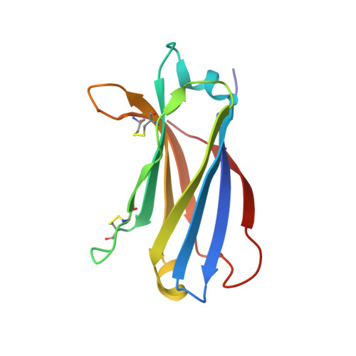
    Kidney- and brain-expressed protein (KIBRA), a multifunctional scaffold protein with around 20 known binding partners, is involved in memory and cognition, organ size control via the Hippo pathway, cell polarity, and membrane trafficking. KIBRA includes tandem N-terminal WW domains, a C2 domain, and motifs for binding atypical PKC and PDZ domains. A naturally occurring human KIBRA variant involving residue changes at positions 734 (Met-to-Ile) and 735 (Ser-to-Ala) within the C2 domain affects cognitive performance. We have elucidated 3D structures and calcium- and phosphoinositide-binding properties of human KIBRA C2 domain. Both WT and variant C2 adopt a canonical type I topology C2 domain fold. Neither Ca 2+ nor any other metal ion was bound to WT or variant KIBRA C2 in crystal structures, and Ca 2+ titration produced no significant reproducible changes in NMR spectra. NMR and X-ray diffraction data indicate that KIBRA C2 binds phosphoinositides via an atypical site involving β-strands 5, 2, 1, and 8. Molecular dynamics simulations indicate that KIBRA C2 interacts with membranes via primary and secondary sites on the same domain face as the experimentally identified phosphoinositide-binding site. Our results indicate that KIBRA C2 domain association with membranes is calcium-independent and involves distinctive C2 domain-membrane relative orientations.


  • Organizational Affiliation
    • From the Department of Biology and Biochemistry, University of Bath, Bath BA2 7AY, United Kingdom.

Macromolecule Content 

  • Total Structure Weight: 29.56 kDa 
  • Atom Count: 2,213 
  • Modeled Residue Count: 258 
  • Deposited Residue Count: 258 
  • Unique protein chains: 1

Macromolecules

Find similar proteins by:|  3D Structure
Entity ID: 1
MoleculeChains  Sequence LengthOrganismDetailsImage
Protein KIBRA
A, B
129Homo sapiensMutation(s): 1 
Gene Names: WWC1KIAA0869
UniProt & NIH Common Fund Data Resources
Find proteins for Q8IX03 (Homo sapiens)
Explore Q8IX03 
Go to UniProtKB:  Q8IX03
PHAROS:  Q8IX03
GTEx:  ENSG00000113645 
Entity Groups
Sequence Clusters30% Identity50% Identity70% Identity90% Identity95% Identity100% Identity
UniProt GroupQ8IX03
Sequence Annotations
Expand
Reference Sequence

Experimental Data & Validation

Experimental Data

  • Method: X-RAY DIFFRACTION
  • Resolution: 2.42 Å
  • R-Value Free:  0.247 (Depositor), 0.248 (DCC) 
  • R-Value Work:  0.201 (Depositor), 0.201 (DCC) 
  • R-Value Observed: 0.203 (Depositor) 
Space Group: P 61 2 2
Unit Cell:
Length ( Å )Angle ( ˚ )
a = 83.88α = 90
b = 83.88β = 90
c = 207.77γ = 120
Software Package:
Software NamePurpose
PHENIXrefinement
d*TREKdata reduction
d*TREKdata scaling
BALBESphasing

Structure Validation

View Full Validation Report


Entry History 

& Funding Information

Deposition Data


Funding OrganizationLocationGrant Number
Biotechnology and Biological Sciences Research CouncilUnited KingdomBB/J008176/1

Revision History  (Full details and data files)

  • Version 1.0: 2018-05-16
    Type: Initial release
  • Version 2.0: 2018-06-27
    Changes: Atomic model, Data collection, Database references
  • Version 2.1: 2024-01-17
    Changes: Data collection, Database references, Refinement description
  • Version 2.2: 2024-11-06
    Changes: Structure summary