6EPX | pdb_00006epx

The ATAD2 bromodomain in complex with compound 3


Experimental Data Snapshot

  • Method: X-RAY DIFFRACTION
  • Resolution: 1.84 Å
  • R-Value Free: 
    0.219 (Depositor), 0.222 (DCC) 
  • R-Value Work: 
    0.205 (Depositor), 0.208 (DCC) 
  • R-Value Observed: 
    0.205 (Depositor) 

wwPDB Validation 3D Report Full Report

Validation slider image for 6EPX

Ligand Structure Quality Assessment 


This is version 1.3 of the entry. See complete history

Literature

Hitting a Moving Target: Simulation and Crystallography Study of ATAD2 Bromodomain Blockers.

Dolbois, A.Batiste, L.Wiedmer, L.Dong, J.Brutsch, M.Huang, D.Deerain, N.M.Spiliotopoulos, D.Cheng-Sanchez, I.Laul, E.Nevado, C.Sledz, P.Caflisch, A.

(2020) ACS Med Chem Lett 11: 1573-1580

  • DOI: https://doi.org/10.1021/acsmedchemlett.0c00080
  • Primary Citation Related Structures: 
    5F36, 6EPJ, 6EPT, 6EPU, 6EPV, 6EPX, 6HI3, 6HI4, 6HI5, 6HI6, 6HI7, 6HI8, 6HIA, 6HIB, 6HIC, 6HID, 6HIE

  • PubMed Abstract: 

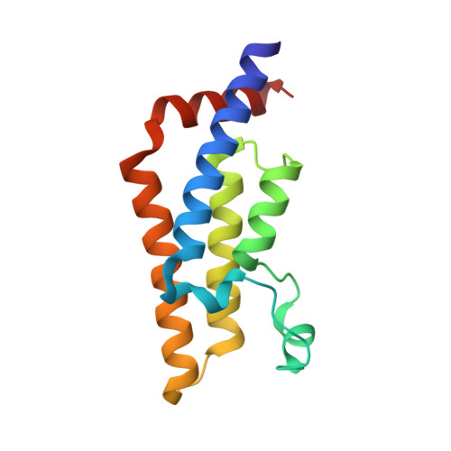
    Small molecule ligand binding to the ATAD2 bromodomain is investigated here through the synergistic combination of molecular dynamics and protein crystallography. A previously unexplored conformation of the binding pocket upon rearrangement of the gatekeeper residue Ile1074 has been found. Further, our investigations reveal how minor structural differences in the ligands result in binding with different plasticity of the ZA loop for this difficult-to-drug bromodomain.


  • Organizational Affiliation
    • Department of Chemistry and Department of Biochemistry, University of Zurich, Winterthurerstrasse 190, 8057 Zurich, Switzerland.

Macromolecule Content 

  • Total Structure Weight: 16.07 kDa 
  • Atom Count: 1,222 
  • Modeled Residue Count: 130 
  • Deposited Residue Count: 130 
  • Unique protein chains: 1

Macromolecules

Find similar proteins by:|  3D Structure
Entity ID: 1
MoleculeChains  Sequence LengthOrganismDetailsImage
ATPase family AAA domain-containing protein 2130Homo sapiensMutation(s): 0 
Gene Names: ATAD2L16PRO2000
EC: 3.6.1.3 (PDB Primary Data), 3.6.1 (UniProt)
UniProt & NIH Common Fund Data Resources
Find proteins for Q6PL18 (Homo sapiens)
Explore Q6PL18 
Go to UniProtKB:  Q6PL18
PHAROS:  Q6PL18
GTEx:  ENSG00000156802 
Entity Groups
Sequence Clusters30% Identity50% Identity70% Identity90% Identity95% Identity100% Identity
UniProt GroupQ6PL18
Sequence Annotations
Expand
Reference Sequence

Experimental Data & Validation

Experimental Data

  • Method: X-RAY DIFFRACTION
  • Resolution: 1.84 Å
  • R-Value Free:  0.219 (Depositor), 0.222 (DCC) 
  • R-Value Work:  0.205 (Depositor), 0.208 (DCC) 
  • R-Value Observed: 0.205 (Depositor) 
Space Group: P 65 2 2
Unit Cell:
Length ( Å )Angle ( ˚ )
a = 79.575α = 90
b = 79.575β = 90
c = 138.187γ = 120
Software Package:
Software NamePurpose
PHENIXrefinement
XDSdata reduction
PHASERphasing

Structure Validation

View Full Validation Report


Ligand Structure Quality Assessment 


Entry History 

Deposition Data

Revision History  (Full details and data files)

  • Version 1.0: 2018-10-31
    Type: Initial release
  • Version 1.1: 2019-11-13
    Changes: Data collection, Database references
  • Version 1.2: 2020-11-04
    Changes: Database references
  • Version 1.3: 2024-05-08
    Changes: Data collection, Database references