6E4R | pdb_00006e4r

Crystal Structure of the Drosophila Melanogaster Polypeptide N-Acetylgalactosaminyl Transferase PGANT9B


Experimental Data Snapshot

  • Method: X-RAY DIFFRACTION
  • Resolution: 2.06 Å
  • R-Value Free: 
    0.207 (Depositor), 0.210 (DCC) 
  • R-Value Work: 
    0.164 (Depositor), 0.168 (DCC) 
  • R-Value Observed: 
    0.167 (Depositor) 

Starting Model: experimental
View more details

wwPDB Validation 3D Report Full Report

Validation slider image for 6E4R

Ligand Structure Quality Assessment 


This is version 1.3 of the entry. See complete history

Literature

A molecular switch orchestrates enzyme specificity and secretory granule morphology.

Ji, S.Samara, N.L.Revoredo, L.Zhang, L.Tran, D.T.Muirhead, K.Tabak, L.A.Ten Hagen, K.G.

(2018) Nat Commun 9: 3508-3508

  • DOI: https://doi.org/10.1038/s41467-018-05978-9
  • Primary Citation Related Structures: 
    6E4Q, 6E4R

  • PubMed Abstract: 

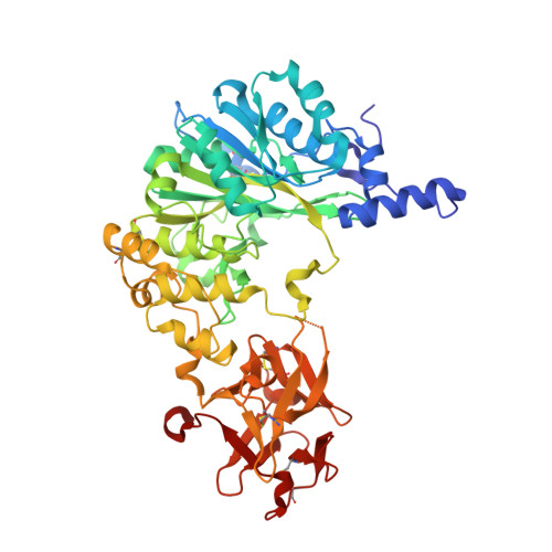
    Regulated secretion is an essential process where molecules destined for export are directed to membranous secretory granules, where they undergo packaging and maturation. Here, we identify a gene (pgant9) that influences the structure and shape of secretory granules within the Drosophila salivary gland. Loss of pgant9, which encodes an O-glycosyltransferase, results in secretory granules with an irregular, shard-like morphology, and altered glycosylation of cargo. Interestingly, pgant9 undergoes a splicing event that acts as a molecular switch to alter the charge of a loop controlling access to the active site of the enzyme. The splice variant with the negatively charged loop glycosylates the positively charged secretory cargo and rescues secretory granule morphology. Our study highlights a mechanism for dictating substrate specificity within the O-glycosyltransferase enzyme family. Moreover, our in vitro and in vivo studies suggest that the glycosylation status of secretory cargo influences the morphology of maturing secretory granules.


  • Organizational Affiliation
    • Developmental Glycobiology Section, NIDCR, National Institutes of Health, 30 Convent Drive, Bethesda, MD, 20892-4370, USA.

Macromolecule Content 

  • Total Structure Weight: 117.64 kDa 
  • Atom Count: 8,921 
  • Modeled Residue Count: 999 
  • Deposited Residue Count: 1,014 
  • Unique protein chains: 1

Macromolecules

Find similar proteins by:|  3D Structure
Entity ID: 1
MoleculeChains  Sequence LengthOrganismDetailsImage
polypeptide N-acetylgalactosaminyltransferase 9
A, B
507Drosophila melanogasterMutation(s): 0 
Gene Names: pgant9CG30463
EC: 2.4.1.41
UniProt
Find proteins for Q8MRC9 (Drosophila melanogaster)
Explore Q8MRC9 
Go to UniProtKB:  Q8MRC9
Entity Groups
Sequence Clusters30% Identity50% Identity70% Identity90% Identity95% Identity100% Identity
UniProt GroupQ8MRC9
Glycosylation
Glycosylation Sites: 1Go to GlyGen: Q8MRC9-1
Sequence Annotations
Expand
Reference Sequence

Small Molecules

Ligands 4 Unique
IDChains Name / Formula / InChI Key2D Diagram3D Interactions
NAG

Query on NAG


Download:Ideal Coordinates CCD File
C [auth A],
L [auth B]
2-acetamido-2-deoxy-beta-D-glucopyranose
C8 H15 N O6
OVRNDRQMDRJTHS-FMDGEEDCSA-N
PGE

Query on PGE


Download:Ideal Coordinates CCD File
I [auth A]TRIETHYLENE GLYCOL
C6 H14 O4
ZIBGPFATKBEMQZ-UHFFFAOYSA-N
GOL

Query on GOL


Download:Ideal Coordinates CCD File
F [auth A]
G [auth A]
H [auth A]
J [auth A]
K [auth A]
F [auth A],
G [auth A],
H [auth A],
J [auth A],
K [auth A],
N [auth B],
O [auth B],
P [auth B]
GLYCEROL
C3 H8 O3
PEDCQBHIVMGVHV-UHFFFAOYSA-N
EDO

Query on EDO


Download:Ideal Coordinates CCD File
D [auth A]
E [auth A]
M [auth B]
Q [auth B]
R [auth B]
D [auth A],
E [auth A],
M [auth B],
Q [auth B],
R [auth B],
S [auth B],
T [auth B]
1,2-ETHANEDIOL
C2 H6 O2
LYCAIKOWRPUZTN-UHFFFAOYSA-N

Experimental Data & Validation

Experimental Data

  • Method: X-RAY DIFFRACTION
  • Resolution: 2.06 Å
  • R-Value Free:  0.207 (Depositor), 0.210 (DCC) 
  • R-Value Work:  0.164 (Depositor), 0.168 (DCC) 
  • R-Value Observed: 0.167 (Depositor) 
Space Group: I 1 2 1
Unit Cell:
Length ( Å )Angle ( ˚ )
a = 113.913α = 90
b = 48.44β = 91.4
c = 232.037γ = 90
Software Package:
Software NamePurpose
PHENIXrefinement
HKL-2000data reduction
Aimlessdata scaling
MOLREPphasing

Structure Validation

View Full Validation Report


Ligand Structure Quality Assessment 


Entry History 

Deposition Data

Revision History  (Full details and data files)

  • Version 1.0: 2018-09-12
    Type: Initial release
  • Version 1.1: 2020-07-29
    Type: Remediation
    Reason: Carbohydrate remediation
    Changes: Data collection, Derived calculations, Structure summary
  • Version 1.2: 2023-10-11
    Changes: Data collection, Database references, Refinement description, Structure summary
  • Version 1.3: 2024-11-06
    Changes: Structure summary