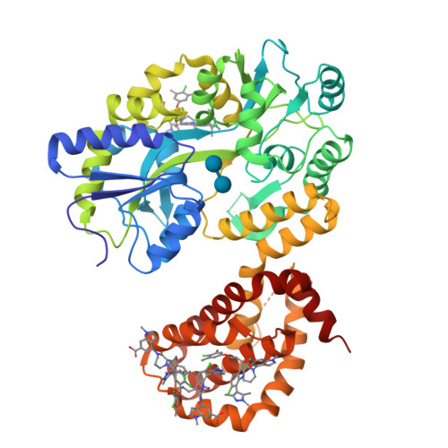
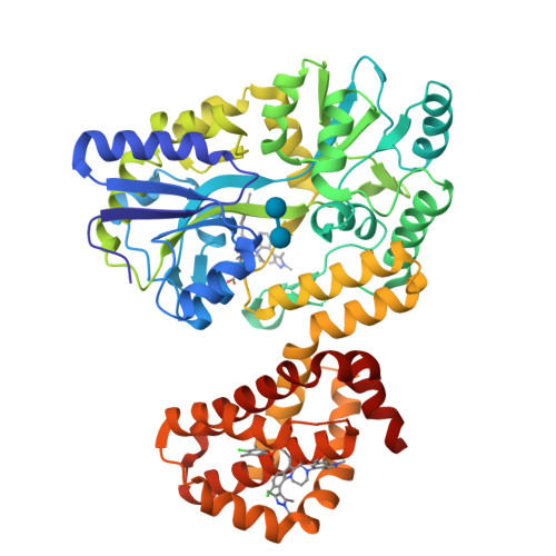
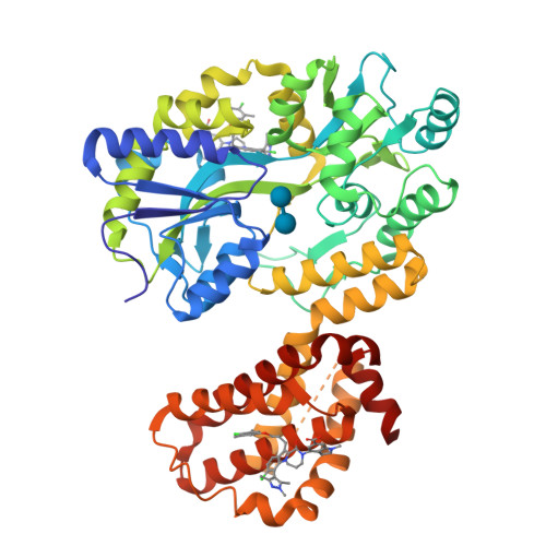
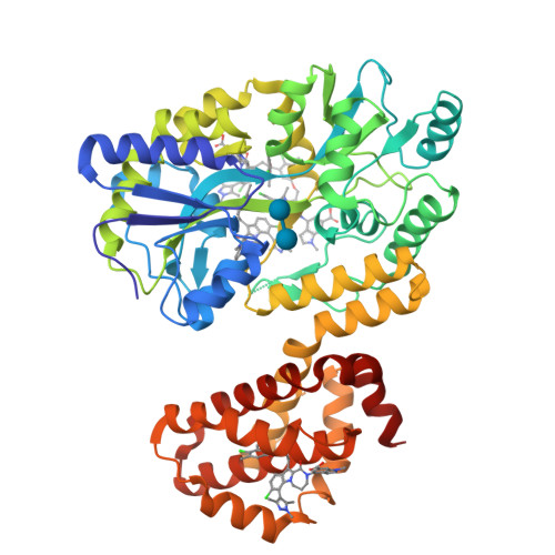
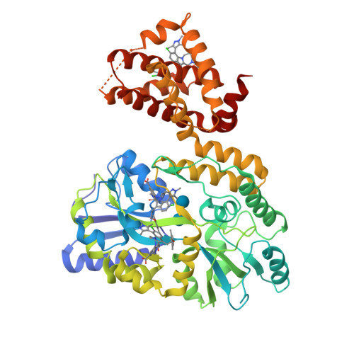
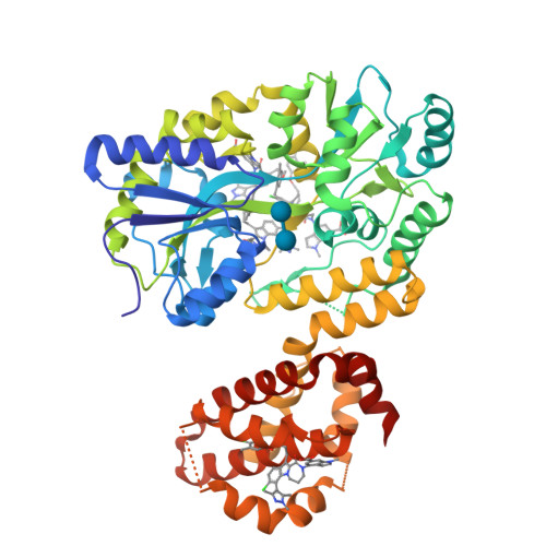
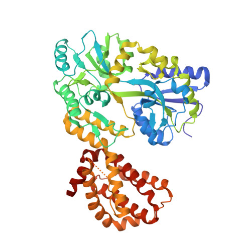
6DM8

Understanding the Species Selectivity of Myeloid cell leukemia-1 (Mcl-1) inhibitors


Experimental Data Snapshot

  • Method: X-RAY DIFFRACTION
  • Resolution: 2.70 Å
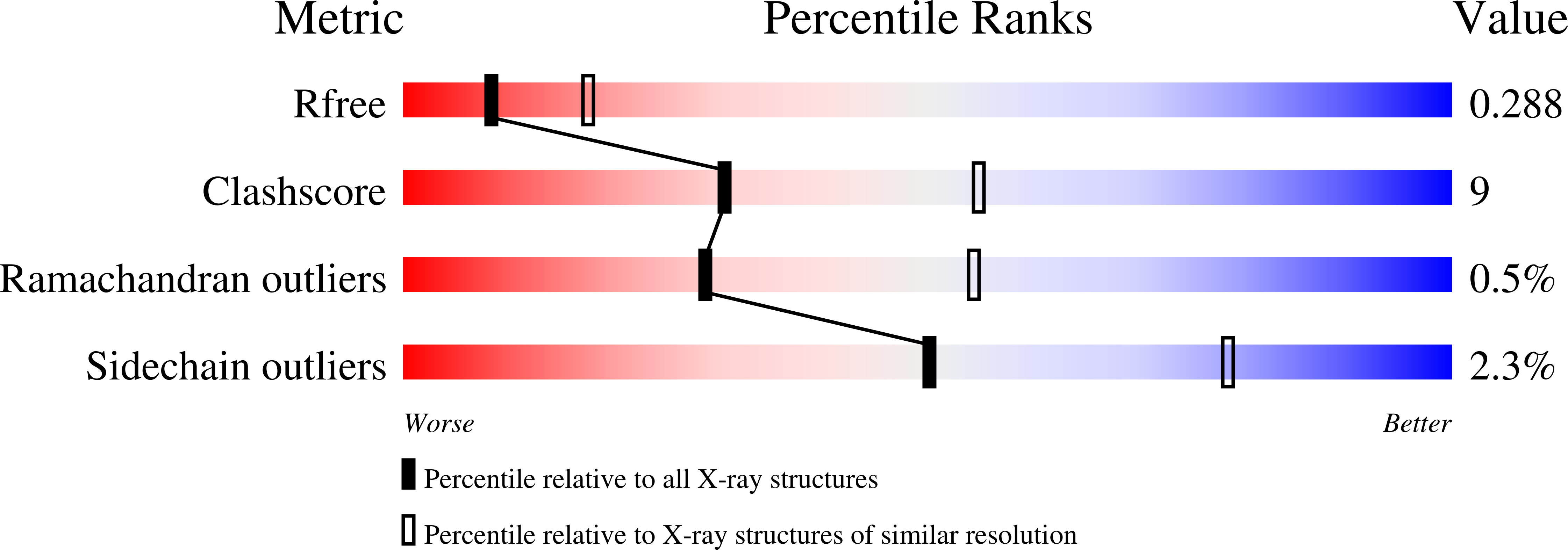
  • R-Value Free: 0.288 
  • R-Value Work: 0.245 
  • R-Value Observed: 0.246 

wwPDB Validation   3D Report Full Report


This is version 2.1 of the entry. See complete history


Literature

Understanding the Species Selectivity of Myeloid Cell Leukemia-1 (Mcl-1) Inhibitors.

Zhao, B.Arnold, A.L.Coronel, M.A.Lee, J.H.Lee, T.Olejniczak, E.T.Fesik, S.W.

(2018) Biochemistry 57: 4952-4958

  • DOI: https://doi.org/10.1021/acs.biochem.8b00626
  • Primary Citation of Related Structures:  
    6DM8

  • PubMed Abstract: 

    To test for on target toxicity of a new chemical entity, it is important to have comparable binding affinities of the compound in the target proteins from humans and the test species. To evaluate our myeloid cell leukemia-1 (Mcl-1) inhibitors, we tested them against rodent Mcl-1 and found a significant loss of binding affinity when compared to that seen with human Mcl-1. To understand the affinity loss, we used sequence alignments and structures of human Mcl-1/inhibitor complexes to identify the important differences in the amino acid sequences. One difference is human L246 (F226 in rat, F227 in mouse) in the ligand binding pocket. Mutating rat F226 to a Leu restores affinity, but the mouse F227L mutant still has a ligand affinity that is lower than that of human Mcl-1. Another mutation of mouse F267, located ∼12 Å from the ligand pocket, to the human/rat cysteine, F267C, improved the affinity and combined with F227L resulted in a mutant mouse protein with a binding affinity similar to that of human Mcl-1. To help understand the structural components of the affinity loss, we obtained an X-ray structure of a mouse Mcl-1/inhibitor complex and identified how the residue changes reduced compound complementarity. Finally, we tested Mcl-1 of other preclinical animal models (canine, monkey, rabbit, and ferret) that are identical to humans in terms of these two residues and found that their Mcl-1 bound our compounds with affinities comparable to that of human Mcl-1. These results have implications for understanding ligand selectivity for similar proteins and for the interpretation of preclinical toxicology studies with Mcl-1 inhibitors.


  • Organizational Affiliation

    Department of Biochemistry , Vanderbilt University School of Medicine , 2215 Garland Avenue, 607 Light Hall , Nashville , Tennessee 37232-0146 , United States.


Macromolecules
Find similar proteins by:  (by identity cutoff)  |  3D Structure
Entity ID: 1
MoleculeChains Sequence LengthOrganismDetailsImage
Induced myeloid leukemia cell differentiation protein Mcl-1 homolog - MBP chimera
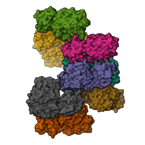
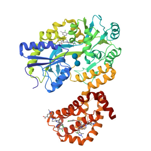
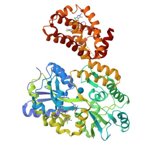
A, B, C, D, E
A, B, C, D, E, F, G, H
518Escherichia coli K-12Mus musculus
This entity is chimeric
Mutation(s): 0 
Gene Names: malEb4034JW3994Mcl1
UniProt & NIH Common Fund Data Resources
Find proteins for P0AEX9 (Escherichia coli (strain K12))
Explore P0AEX9 
Go to UniProtKB:  P0AEX9
Find proteins for P97287 (Mus musculus)
Explore P97287 
Go to UniProtKB:  P97287
IMPC:  MGI:101769
Entity Groups  
Sequence Clusters30% Identity50% Identity70% Identity90% Identity95% Identity100% Identity
UniProt GroupsP97287P0AEX9
Sequence Annotations
Expand
  • Reference Sequence
Oligosaccharides

Help

Entity ID: 2
MoleculeChains Length2D Diagram Glycosylation3D Interactions
alpha-D-glucopyranose-(1-4)-alpha-D-glucopyranose
I, J, K, L, M
I, J, K, L, M, N, O, P
2N/A
Glycosylation Resources
GlyTouCan:  G07411ON
GlyCosmos:  G07411ON
Small Molecules
Ligands 1 Unique
IDChains Name / Formula / InChI Key2D Diagram3D Interactions
6AK
Query on 6AK

Download Ideal Coordinates CCD File 
AA [auth D]
BA [auth E]
CA [auth E]
DA [auth F]
EA [auth F]
AA [auth D],
BA [auth E],
CA [auth E],
DA [auth F],
EA [auth F],
FA [auth F],
GA [auth F],
HA [auth G],
IA [auth G],
JA [auth G],
KA [auth H],
LA [auth H],
MA [auth H],
NA [auth H],
Q [auth A],
R [auth A],
S [auth A],
T [auth B],
U [auth B],
V [auth C],
W [auth C],
X [auth C],
Y [auth C],
Z [auth D]
4-{8-chloro-11-[3-(4-chloro-3,5-dimethylphenoxy)propyl]-1-oxo-7-(1,3,5-trimethyl-1H-pyrazol-4-yl)-4,5-dihydro-1H-[1,4]diazepino[1,2-a]indol-2(3H)-yl}-1-methyl-1H-indole-6-carboxylic acid
C39 H39 Cl2 N5 O4
BDINUPBROUJUMH-UHFFFAOYSA-N
Binding Affinity Annotations 
IDSourceBinding Affinity
6AK BindingDB:  6DM8 Ki: min: 0.5, max: 50 (nM) from 7 assay(s)
Experimental Data & Validation

Experimental Data

  • Method: X-RAY DIFFRACTION
  • Resolution: 2.70 Å
  • R-Value Free: 0.288 
  • R-Value Work: 0.245 
  • R-Value Observed: 0.246 
  • Space Group: P 1
Unit Cell:
Length ( Å )Angle ( ˚ )
a = 76.206α = 89.86
b = 104.153β = 89.8
c = 146.206γ = 89.97
Software Package:
Software NamePurpose
PHENIXrefinement
HKL-2000data reduction
HKL-2000data scaling
PHASERphasing

Structure Validation

View Full Validation Report


Entry History & Funding Information

Deposition Data

  • Released Date: 2018-08-01 
  • Deposition Author(s): Zhao, B.

Funding OrganizationLocationGrant Number
National Institutes of Health/National Cancer Institute (NIH/NCI)United StatesBOA 29XS129TO22

Revision History  (Full details and data files)

  • Version 1.0: 2018-08-01
    Type: Initial release
  • Version 1.1: 2018-08-22
    Changes: Data collection, Database references
  • Version 1.2: 2019-12-04
    Changes: Author supporting evidence
  • Version 2.0: 2020-07-29
    Type: Remediation
    Reason: Carbohydrate remediation
    Changes: Atomic model, Data collection, Derived calculations, Non-polymer description, Structure summary
  • Version 2.1: 2024-03-13
    Changes: Advisory, Data collection, Database references, Structure summary