6DID | pdb_00006did

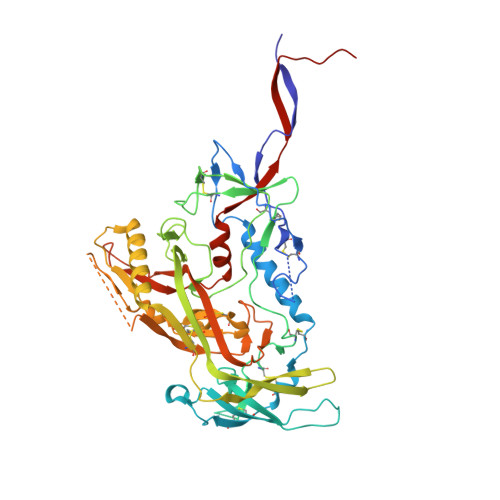
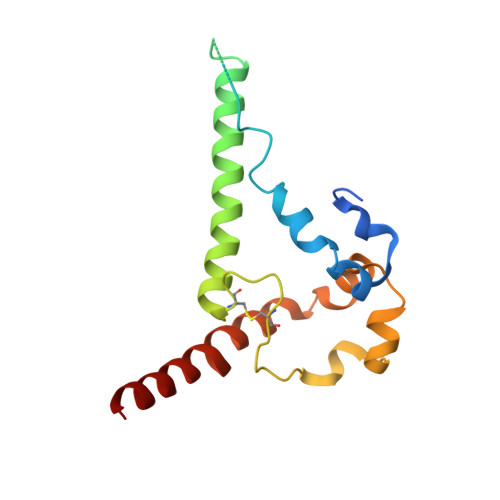
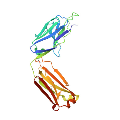
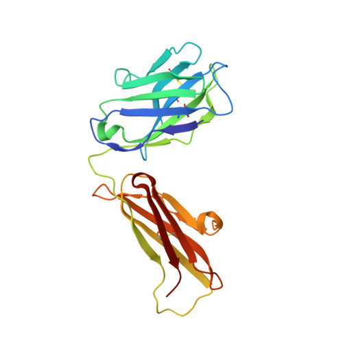
HIV Env BG505 SOSIP with polyclonal Fabs from immunized rabbit #3417 post-boost#1


Experimental Data Snapshot

  • Method: ELECTRON MICROSCOPY
  • Resolution: 4.71 Å
  • Aggregation State: PARTICLE 
  • Reconstruction Method: SINGLE PARTICLE 

wwPDB Validation 3D Report Full Report

Validation slider image for 6DID

This is version 2.1 of the entry. See complete history. 

Literature

Electron-Microscopy-Based Epitope Mapping Defines Specificities of Polyclonal Antibodies Elicited during HIV-1 BG505 Envelope Trimer Immunization.

Bianchi, M., Turner, H.L., Nogal, B., Cottrell, C.A., Oyen, D., Pauthner, M., Bastidas, R., Nedellec, R., McCoy, L.E., Wilson, I.A., Burton, D.R., Ward, A.B., Hangartner, L.

(2018) Immunity 49: 288-300.e8

  • DOI: https://doi.org/10.1016/j.immuni.2018.07.009
  • Primary Citation Related Structures: 
    6CJK, 6DID

  • PubMed Abstract: 

    Characterizing polyclonal antibody responses via currently available methods is inherently complex and difficult. Mapping epitopes in an immune response is typically incomplete, which creates a barrier to fully understanding the humoral response to antigens and hinders rational vaccine design efforts. Here, we describe a method of characterizing polyclonal responses by using electron microscopy, and we applied this method to the immunization of rabbits with an HIV-1 envelope glycoprotein vaccine candidate, BG505 SOSIP.664. We detected known epitopes within the polyclonal sera and revealed how antibody responses evolved during the prime-boosting strategy to ultimately result in a neutralizing antibody response. We uncovered previously unidentified epitopes, including an epitope proximal to one recognized by human broadly neutralizing antibodies as well as potentially distracting non-neutralizing epitopes. Our method provides an efficient and semiquantitative map of epitopes that are targeted in a polyclonal antibody response and should be of widespread utility in vaccine and infection studies.


  • Organizational Affiliation: 

    • Department of Immunology and Microbiology, The Scripps Research Institute, La Jolla, CA 92037, USA.

Macromolecule Content 

  • Total Structure Weight: 378.24 kDa 
  • Atom Count: 25,209 
  • Modeled Residue Count: 3,009 
  • Deposited Residue Count: 3,195 
  • Unique protein chains: 4

Macromolecules

Find similar proteins by:|  3D Structure
Entity ID: 1
MoleculeChains  Sequence LengthOrganismDetailsImage
Envelope glycoprotein gp160A,
C [auth F],
E [auth G]
481Human immunodeficiency virus 1Mutation(s): 0 
Gene Names: env
UniProt
Find proteins for Q2N0S6 (Human immunodeficiency virus type 1)
Explore Q2N0S6 
Go to UniProtKB:  Q2N0S6
Entity Groups
Sequence Clusters30% Identity50% Identity70% Identity90% Identity95% Identity100% Identity
UniProt GroupQ2N0S6
Glycosylation
Glycosylation Sites: 17
Sequence Annotations
Expand
Reference Sequence
Find similar proteins by:|  3D Structure
Entity ID: 2
MoleculeChains  Sequence LengthOrganismDetailsImage
Envelope glycoprotein gp160B,
D [auth I],
F [auth J]
153Human immunodeficiency virus 1Mutation(s): 0 
Gene Names: env
UniProt
Find proteins for Q2N0S7 (Human immunodeficiency virus type 1)
Explore Q2N0S7 
Go to UniProtKB:  Q2N0S7
Entity Groups
Sequence Clusters30% Identity50% Identity70% Identity90% Identity95% Identity100% Identity
UniProt GroupQ2N0S7
Glycosylation
Glycosylation Sites: 2
Sequence Annotations
Expand
Reference Sequence
Find similar proteins by:|  3D Structure
Entity ID: 3
MoleculeChains  Sequence LengthOrganismDetailsImage
Monoclonal antibody 10A light chainG [auth C],
I [auth E],
K [auth H]
217Oryctolagus cuniculusMutation(s): 0 
UniProt
Find proteins for P01840 (Oryctolagus cuniculus)
Explore P01840 
Go to UniProtKB:  P01840
Entity Groups
Sequence Clusters30% Identity50% Identity70% Identity90% Identity95% Identity100% Identity
UniProt GroupP01840
Sequence Annotations
Expand
Reference Sequence
Find similar proteins by:|  3D Structure
Entity ID: 4
MoleculeChains  Sequence LengthOrganismDetailsImage
Monoclonal antibody 10A heavy chainH [auth D],
J [auth K],
L
214Oryctolagus cuniculusMutation(s): 0 
UniProt
Find proteins for A0A1Y1B8B1 (Oryctolagus cuniculus)
Explore A0A1Y1B8B1 
Go to UniProtKB:  A0A1Y1B8B1
Entity Groups
Sequence Clusters30% Identity50% Identity70% Identity90% Identity95% Identity100% Identity
UniProt GroupA0A1Y1B8B1
Sequence Annotations
Expand
Reference Sequence

Oligosaccharides

Help  
Entity ID: 5
MoleculeChains Length2D Diagram GlycosylationD Interactions
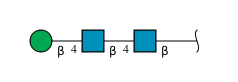
beta-D-mannopyranose-(1-4)-2-acetamido-2-deoxy-beta-D-glucopyranose-(1-4)-2-acetamido-2-deoxy-beta-D-glucopyranoseKA [auth k],
M,
Y
3N-Glycosylation
Glycosylation Resources
GlyTouCan: G15407YE
GlyCosmos: G15407YE
GlyGen: G15407YE
Entity ID: 7
MoleculeChains Length2D Diagram GlycosylationD Interactions
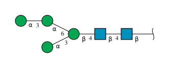
alpha-D-mannopyranose-(1-2)-alpha-D-mannopyranose-(1-3)-[alpha-D-mannopyranose-(1-3)-alpha-D-mannopyranose-(1-6)]beta-D-mannopyranose-(1-4)-2-acetamido-2-deoxy-beta-D-glucopyranose-(1-4)-2-acetamido-2-deoxy-beta-D-glucopyranoseCA [auth c],
OA [auth o],
Q
7N-Glycosylation
Glycosylation Resources
GlyTouCan: G07617FP
GlyCosmos: G07617FP
GlyGen: G07617FP
Entity ID: 8
MoleculeChains Length2D Diagram GlycosylationD Interactions
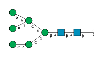
alpha-D-mannopyranose-(1-2)-alpha-D-mannopyranose-(1-3)-[alpha-D-mannopyranose-(1-3)-[alpha-D-mannopyranose-(1-6)]alpha-D-mannopyranose-(1-6)]beta-D-mannopyranose-(1-4)-2-acetamido-2-deoxy-beta-D-glucopyranose-(1-4)-2-acetamido-2-deoxy-beta-D-glucopyranoseDA [auth d],
PA [auth p],
R
8N-Glycosylation
Glycosylation Resources
GlyTouCan: G80966KZ
GlyCosmos: G80966KZ
GlyGen: G80966KZ
Entity ID: 9
MoleculeChains Length2D Diagram GlycosylationD Interactions
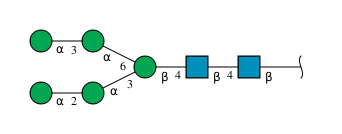
alpha-D-mannopyranose-(1-3)-alpha-D-mannopyranose-(1-6)-[alpha-D-mannopyranose-(1-3)]beta-D-mannopyranose-(1-4)-2-acetamido-2-deoxy-beta-D-glucopyranose-(1-4)-2-acetamido-2-deoxy-beta-D-glucopyranoseHA [auth h],
TA [auth t],
V
6N-Glycosylation
Glycosylation Resources
GlyTouCan: G09724ZC
GlyCosmos: G09724ZC
GlyGen: G09724ZC
Entity ID: 10
MoleculeChains Length2D Diagram GlycosylationD Interactions
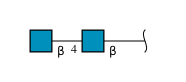
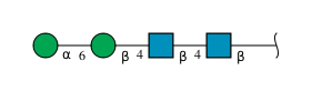
alpha-D-mannopyranose-(1-6)-beta-D-mannopyranose-(1-4)-2-acetamido-2-deoxy-beta-D-glucopyranose-(1-4)-2-acetamido-2-deoxy-beta-D-glucopyranoseIA [auth i],
UA [auth u],
W
4N-Glycosylation
Glycosylation Resources
GlyTouCan: G22573RC
GlyCosmos: G22573RC
GlyGen: G22573RC

Small Molecules

Ligands 1 Unique
IDChains Name / Formula / InChI Key2D Diagram3D Interactions
NAG

Query on NAG


Download:Ideal Coordinates CCD File
AB [auth A]
BB [auth B]
CB [auth B]
DB [auth F]
EB [auth F]
AB [auth A],
BB [auth B],
CB [auth B],
DB [auth F],
EB [auth F],
FB [auth F],
GB [auth F],
HB [auth F],
IB [auth I],
JB [auth I],
KB [auth G],
LB [auth G],
MB [auth G],
NB [auth G],
OB [auth G],
PB [auth J],
QB [auth J],
WA [auth A],
XA [auth A],
YA [auth A],
ZA [auth A]
2-acetamido-2-deoxy-beta-D-glucopyranose
C8 H15 N O6
OVRNDRQMDRJTHS-FMDGEEDCSA-N

Experimental Data & Validation

Experimental Data

  • Method: ELECTRON MICROSCOPY
  • Resolution: 4.71 Å
  • Aggregation State: PARTICLE 
  • Reconstruction Method: SINGLE PARTICLE 
EM Software:
TaskSoftware PackageVersion
RECONSTRUCTIONcryoSPARC

Structure Validation

View Full Validation Report


Entry History 

& Funding Information

Deposition Data


Funding OrganizationLocationGrant Number
National Institutes of Health/National Institute Of Allergy and Infectious Diseases (NIH/NIAID)United StatesUM1 AI100663
Bill & Melinda Gates FoundationUnited StatesOPP1115782

Revision History  (Full details and data files)

  • Version 1.0: 2018-09-05
    Type: Initial release
  • Version 1.1: 2019-12-18
    Changes: Author supporting evidence
  • Version 2.0: 2020-07-29
    Type: Remediation
    Reason: Carbohydrate remediation
    Changes: Advisory, Atomic model, Data collection, Derived calculations, Structure summary
  • Version 2.1: 2024-11-20
    Changes: Data collection, Database references, Derived calculations, Structure summary