6CJK | pdb_00006cjk

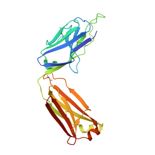
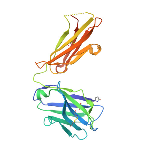
Anti HIV Fab 10A


Experimental Data Snapshot

  • Method: X-RAY DIFFRACTION
  • Resolution: 1.79 Å
  • R-Value Free: 
    0.206 (Depositor), 0.206 (DCC) 
  • R-Value Work: 
    0.163 (Depositor), 0.164 (DCC) 
  • R-Value Observed: 
    0.166 (Depositor) 

wwPDB Validation 3D Report Full Report

Validation slider image for 6CJK

This is version 2.1 of the entry. See complete history. 

Literature

Electron-Microscopy-Based Epitope Mapping Defines Specificities of Polyclonal Antibodies Elicited during HIV-1 BG505 Envelope Trimer Immunization.

Bianchi, M., Turner, H.L., Nogal, B., Cottrell, C.A., Oyen, D., Pauthner, M., Bastidas, R., Nedellec, R., McCoy, L.E., Wilson, I.A., Burton, D.R., Ward, A.B., Hangartner, L.

(2018) Immunity 49: 288-300.e8

  • DOI: https://doi.org/10.1016/j.immuni.2018.07.009
  • Primary Citation Related Structures: 
    6CJK, 6DID

  • PubMed Abstract: 

    Characterizing polyclonal antibody responses via currently available methods is inherently complex and difficult. Mapping epitopes in an immune response is typically incomplete, which creates a barrier to fully understanding the humoral response to antigens and hinders rational vaccine design efforts. Here, we describe a method of characterizing polyclonal responses by using electron microscopy, and we applied this method to the immunization of rabbits with an HIV-1 envelope glycoprotein vaccine candidate, BG505 SOSIP.664. We detected known epitopes within the polyclonal sera and revealed how antibody responses evolved during the prime-boosting strategy to ultimately result in a neutralizing antibody response. We uncovered previously unidentified epitopes, including an epitope proximal to one recognized by human broadly neutralizing antibodies as well as potentially distracting non-neutralizing epitopes. Our method provides an efficient and semiquantitative map of epitopes that are targeted in a polyclonal antibody response and should be of widespread utility in vaccine and infection studies.


  • Organizational Affiliation: 
    • Department of Immunology and Microbiology, The Scripps Research Institute, La Jolla, CA 92037, USA.

Macromolecule Content 

  • Total Structure Weight: 93.1 kDa 
  • Atom Count: 6,980 
  • Modeled Residue Count: 855 
  • Deposited Residue Count: 880 
  • Unique protein chains: 2

Macromolecules

Find similar proteins by:|  3D Structure
Entity ID: 1
MoleculeChains  Sequence LengthOrganismDetailsImage
Immunoglobulin Fab light chain
A, C
217Oryctolagus cuniculusMutation(s): 0 
Entity Groups
Sequence Clusters30% Identity50% Identity70% Identity90% Identity95% Identity100% Identity
Sequence Annotations
Expand
Reference Sequence
Find similar proteins by:|  3D Structure
Entity ID: 2
MoleculeChains  Sequence LengthOrganismDetailsImage
Immunoglobulin Fab heavy chain
B, D
223Oryctolagus cuniculusMutation(s): 0 
Entity Groups
Sequence Clusters30% Identity50% Identity70% Identity90% Identity95% Identity100% Identity
Sequence Annotations
Expand
Reference Sequence

Experimental Data & Validation

Experimental Data

  • Method: X-RAY DIFFRACTION
  • Resolution: 1.79 Å
  • R-Value Free:  0.206 (Depositor), 0.206 (DCC) 
  • R-Value Work:  0.163 (Depositor), 0.164 (DCC) 
  • R-Value Observed: 0.166 (Depositor) 
Space Group: P 1
Unit Cell:
Length ( Å )Angle ( ˚ )
a = 51.173α = 67.99
b = 63.558β = 87.87
c = 72.649γ = 76.73
Software Package:
Software NamePurpose
PHENIXrefinement
HKL-2000data reduction
SCALEPACKdata scaling
PHASERphasing

Structure Validation

View Full Validation Report


Entry History 

& Funding Information

Deposition Data


Funding OrganizationLocationGrant Number
International AIDS Vaccine InitiativeIAVI SFP 2120/2121
National Institutes of Health/National Institute Of Allergy and Infectious Diseases (NIH/NIAID)United StatesUM1AI100663

Revision History  (Full details and data files)

  • Version 1.0: 2018-08-15
    Type: Initial release
  • Version 1.1: 2018-08-29
    Changes: Data collection, Database references
  • Version 2.0: 2019-12-18
    Changes: Author supporting evidence, Polymer sequence
  • Version 2.1: 2024-11-06
    Changes: Advisory, Data collection, Database references, Structure summary