6CUG

Crystal structure of BC8B TCR-CD1b-PC complex


Experimental Data Snapshot

  • Method: X-RAY DIFFRACTION
  • Resolution: 2.40 Å
  • R-Value Free: 
    0.229 (Depositor), 0.250 (DCC) 
  • R-Value Work: 
    0.187 (Depositor), 0.200 (DCC) 
  • R-Value Observed: 
    0.189 (Depositor) 

Starting Models: experimental
View more details

wwPDB Validation   3D Report Full Report


Ligand Structure Quality Assessment 

Created with Raphaël 2.3.0Worse 01 BetterLigand structure goodness of fit to experimental dataBest fitted POVClick on this verticalbar to view detailsBest fitted CUYClick on this verticalbar to view detailsBest fitted NAGClick on this verticalbar to view details

This is version 1.4 of the entry. See complete history


Literature

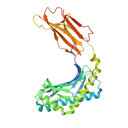
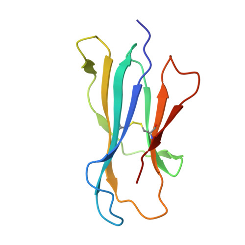
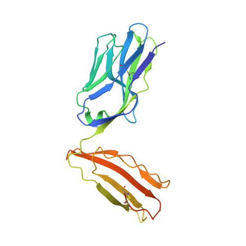
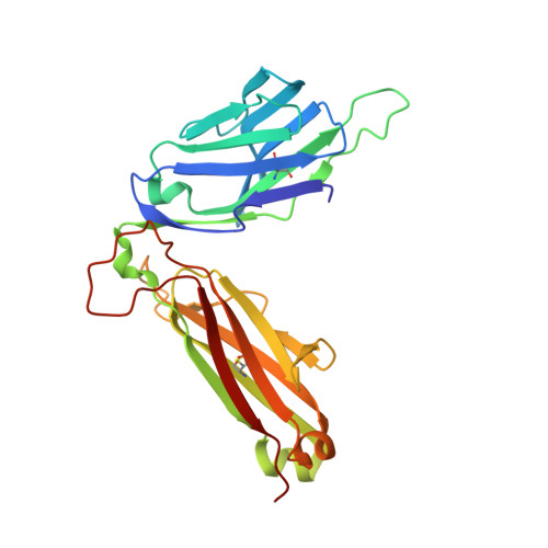
A T-cell receptor escape channel allows broad T-cell response to CD1b and membrane phospholipids.

Shahine, A.Reinink, P.Reijneveld, J.F.Gras, S.Holzheimer, M.Cheng, T.Y.Minnaard, A.J.Altman, J.D.Lenz, S.Prandi, J.Kubler-Kielb, J.Moody, D.B.Rossjohn, J.Van Rhijn, I.

(2019) Nat Commun 10: 56-56

  • DOI: https://doi.org/10.1038/s41467-018-07898-0
  • Primary Citation of Related Structures:  
    6CUG, 6CUH, 6D64

  • PubMed Abstract: 

    CD1 proteins are expressed on dendritic cells, where they display lipid antigens to T-cell receptors (TCRs). Here we describe T-cell autoreactivity towards ubiquitous human membrane phospholipids presented by CD1b. These T-cells discriminate between two major types of lipids, sphingolipids and phospholipids, but were broadly cross-reactive towards diverse phospholipids including phosphatidylcholine, phosphatidylinositol and phosphatidylethanolamine. The crystal structure of a representative TCR bound to CD1b-phosphatidylcholine provides a molecular mechanism for this promiscuous recognition. We observe a lateral escape channel in the TCR, which shunted phospholipid head groups sideways along the CD1b-TCR interface, without contacting the TCR. Instead the TCR recognition site involved the neck region phosphate that is common to all major self-phospholipids but absent in sphingolipids. Whereas prior studies have focused on foreign lipids or rare self-lipids, we define a new molecular mechanism of promiscuous recognition of common self-phospholipids including those that are known targets in human autoimmune disease.


  • Organizational Affiliation

    Infection and Immunity Program and Department of Biochemistry and Molecular Biology, Biomedicine Discovery Institute, Monash University, Clayton, VIC, 3800, Australia.


Macromolecules
Find similar proteins by:  (by identity cutoff)  |  3D Structure
Entity ID: 1
MoleculeChains Sequence LengthOrganismDetailsImage
T-cell surface glycoprotein CD1b300Homo sapiensMutation(s): 0 
Gene Names: CD1B
UniProt & NIH Common Fund Data Resources
Find proteins for P29016 (Homo sapiens)
Explore P29016 
Go to UniProtKB:  P29016
PHAROS:  P29016
GTEx:  ENSG00000158485 
Entity Groups  
Sequence Clusters30% Identity50% Identity70% Identity90% Identity95% Identity100% Identity
UniProt GroupP29016
Glycosylation
Glycosylation Sites: 1Go to GlyGen: P29016-1
Sequence Annotations
Expand
  • Reference Sequence
Find similar proteins by:  (by identity cutoff)  |  3D Structure
Entity ID: 2
MoleculeChains Sequence LengthOrganismDetailsImage
Beta-2-microglobulin99Homo sapiensMutation(s): 0 
Gene Names: B2MCDABP0092HDCMA22P
UniProt & NIH Common Fund Data Resources
Find proteins for P61769 (Homo sapiens)
Explore P61769 
Go to UniProtKB:  P61769
PHAROS:  P61769
GTEx:  ENSG00000166710 
Entity Groups  
Sequence Clusters30% Identity50% Identity70% Identity90% Identity95% Identity100% Identity
UniProt GroupP61769
Sequence Annotations
Expand
  • Reference Sequence
Find similar proteins by:  (by identity cutoff)  |  3D Structure
Entity ID: 3
MoleculeChains Sequence LengthOrganismDetailsImage
T-cell receptor alpha variable TRAV9-2 - BC8B TCRC [auth D]207Homo sapiensMutation(s): 0 
Entity Groups  
Sequence Clusters30% Identity50% Identity70% Identity90% Identity95% Identity100% Identity
Sequence Annotations
Expand
  • Reference Sequence
Find similar proteins by:  (by identity cutoff)  |  3D Structure
Entity ID: 4
MoleculeChains Sequence LengthOrganismDetailsImage
T-cell receptor beta variable TRBV6-2 - BC8B TCRD [auth E]245Homo sapiensMutation(s): 0 
Entity Groups  
Sequence Clusters30% Identity50% Identity70% Identity90% Identity95% Identity100% Identity
Sequence Annotations
Expand
  • Reference Sequence
Experimental Data & Validation

Experimental Data

  • Method: X-RAY DIFFRACTION
  • Resolution: 2.40 Å
  • R-Value Free:  0.229 (Depositor), 0.250 (DCC) 
  • R-Value Work:  0.187 (Depositor), 0.200 (DCC) 
  • R-Value Observed: 0.189 (Depositor) 
Space Group: P 1 21 1
Unit Cell:
Length ( Å )Angle ( ˚ )
a = 73.3α = 90
b = 65.7β = 100.7
c = 101.9γ = 90
Software Package:
Software NamePurpose
BUSTERrefinement
XDSdata reduction
SCALAdata scaling
PHASERphasing
PDB_EXTRACTdata extraction

Structure Validation

View Full Validation Report


Ligand Structure Quality Assessment 

Created with Raphaël 2.3.0Worse 01 BetterLigand structure goodness of fit to experimental dataBest fitted POVClick on this verticalbar to view detailsBest fitted CUYClick on this verticalbar to view detailsBest fitted NAGClick on this verticalbar to view details

Entry History 

Deposition Data

Revision History  (Full details and data files)

  • Version 1.0: 2019-01-16
    Type: Initial release
  • Version 1.1: 2020-07-29
    Type: Remediation
    Reason: Carbohydrate remediation
    Changes: Data collection, Derived calculations, Structure summary
  • Version 1.2: 2023-10-04
    Changes: Data collection, Database references, Refinement description, Structure summary
  • Version 1.3: 2024-06-05
    Changes: Data collection
  • Version 1.4: 2024-10-30
    Changes: Structure summary