6CLW | pdb_00006clw

Crystal structure of TnmH


Experimental Data Snapshot

  • Method: X-RAY DIFFRACTION
  • Resolution: 2.74 Å
  • R-Value Free: 
    0.254 (Depositor), 0.256 (DCC) 
  • R-Value Work: 
    0.202 (Depositor), 0.207 (DCC) 
  • R-Value Observed: 
    0.205 (Depositor) 

Starting Model: experimental
View more details

wwPDB Validation 3D Report Full Report

Validation slider image for 6CLW

This is version 1.3 of the entry. See complete history

Literature

Characterization of TnmH as anO-Methyltransferase Revealing Insights into Tiancimycin Biosynthesis and Enabling a Biocatalytic Strategy To Prepare Antibody-Tiancimycin Conjugates.

Adhikari, A.Teijaro, C.N.Yan, X.Chang, C.Y.Gui, C.Liu, Y.C.Crnovcic, I.Yang, D.Annaval, T.Rader, C.Shen, B.

(2020) J Med Chem 63: 8432-8441

  • DOI: https://doi.org/10.1021/acs.jmedchem.0c00799
  • Primary Citation Related Structures: 
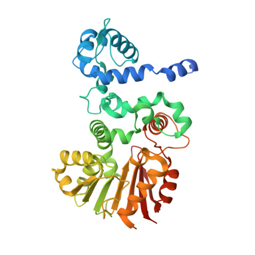
    6CLW, 6CLX

  • PubMed Abstract: 

    The enediynes are among the most cytotoxic molecules known, and their use as anticancer drugs has been successfully demonstrated by targeted delivery. Clinical advancement of the anthraquinone-fused enediynes has been hindered by their low titers and lack of functional groups to enable the preparation of antibody-drug conjugates (ADCs). Here we report biochemical and structural characterization of TnmH from the tiancimycin (TNM) biosynthetic pathway, revealing that (i) TnmH catalyzes regiospecific methylation at the C-7 hydroxyl group, (ii) TnmH exhibits broad substrate promiscuity toward hydroxyanthraquinones and S-alkylated SAM analogues and catalyzes efficient installation of reactive alkyl handles, (iii) the X-ray crystal structure of TnmH provides the molecular basis to account for its broad substrate promiscuity, and (iv) TnmH as a biocatalyst enables the development of novel conjugation strategies to prepare antibody-TNM conjugates. These findings should greatly facilitate the construction and evaluation of antibody-TNM conjugates as next-generation ADCs for targeted chemotherapy.

Macromolecule Content 

  • Total Structure Weight: 77.13 kDa 
  • Atom Count: 5,116 
  • Modeled Residue Count: 680 
  • Deposited Residue Count: 728 
  • Unique protein chains: 1

Macromolecules

Find similar proteins by:|  3D Structure
Entity ID: 1
MoleculeChains  Sequence LengthOrganismDetailsImage
O-methyltransferase
A, B
364Streptomyces sp. CB03234Mutation(s): 0 
Gene Names: tnmHAMK26_31990
UniProt
Find proteins for A0A125SA05 (Streptomyces sp. (strain CB03234))
Explore A0A125SA05 
Go to UniProtKB:  A0A125SA05
Entity Groups
Sequence Clusters30% Identity50% Identity70% Identity90% Identity95% Identity100% Identity
UniProt GroupA0A125SA05
Sequence Annotations
Expand
Reference Sequence

Experimental Data & Validation

Experimental Data

  • Method: X-RAY DIFFRACTION
  • Resolution: 2.74 Å
  • R-Value Free:  0.254 (Depositor), 0.256 (DCC) 
  • R-Value Work:  0.202 (Depositor), 0.207 (DCC) 
  • R-Value Observed: 0.205 (Depositor) 
Space Group: P 31 2 1
Unit Cell:
Length ( Å )Angle ( ˚ )
a = 110.86α = 90
b = 110.86β = 90
c = 113.31γ = 120
Software Package:
Software NamePurpose
REFMACrefinement
MOSFLMdata reduction
SCALAdata scaling
MOLREPphasing

Structure Validation

View Full Validation Report


Entry History 

Deposition Data

Revision History  (Full details and data files)

  • Version 1.0: 2019-03-06
    Type: Initial release
  • Version 1.1: 2019-12-04
    Changes: Author supporting evidence
  • Version 1.2: 2020-09-16
    Changes: Database references
  • Version 1.3: 2023-10-04
    Changes: Data collection, Database references, Refinement description