6CDN | pdb_00006cdn

Crystal structure of cysteine-bound ferrous form of the crosslinked Cl-Tyr157 human cysteine dioxygenase

  • Classification: OXIDOREDUCTASE
  • Organism(s): Homo sapiens
  • Expression System: Escherichia coli
  • Mutation(s): No 

  • Deposited: 2018-02-08 Released: 2018-07-04 
  • Deposition Author(s): Liu, A., Li, J., Shin, I.
  • Funding Organization(s): National Science Foundation (NSF, United States), National Institutes of Health/National Institute of General Medical Sciences (NIH/NIGMS)

Experimental Data Snapshot

  • Method: X-RAY DIFFRACTION
  • Resolution: 2.06 Å
  • R-Value Free: 
    0.193 (Depositor), 0.194 (DCC) 
  • R-Value Work: 
    0.162 (Depositor), 0.162 (DCC) 
  • R-Value Observed: 
    0.165 (Depositor) 

Starting Model: experimental
View more details

wwPDB Validation 3D Report Full Report

Validation slider image for 6CDN

Ligand Structure Quality Assessment 


This is version 1.5 of the entry. See complete history

Literature

Cleavage of a carbon-fluorine bond by an engineered cysteine dioxygenase.

Li, J.Griffith, W.P.Davis, I.Shin, I.Wang, J.Li, F.Wang, Y.Wherritt, D.J.Liu, A.

(2018) Nat Chem Biol 14: 853-860

  • DOI: https://doi.org/10.1038/s41589-018-0085-5
  • Primary Citation Related Structures: 
    6BGF, 6BGM, 6BPS, 6BPT, 6BPU, 6BPV, 6BPW, 6BPX, 6CDH, 6CDN

  • PubMed Abstract: 

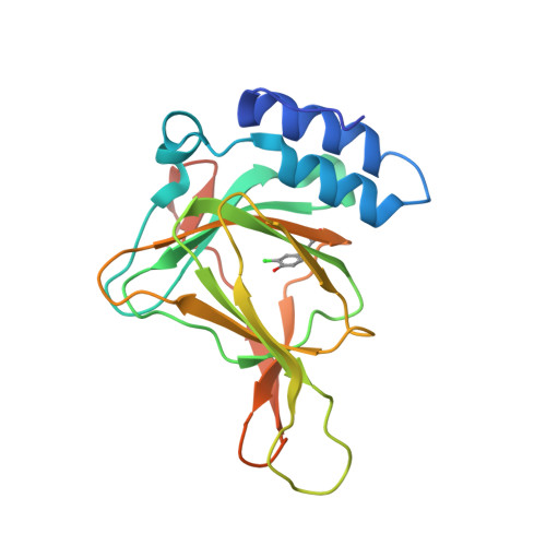
    Cysteine dioxygenase (CDO) plays an essential role in sulfur metabolism by regulating homeostatic levels of cysteine. Human CDO contains a post-translationally generated Cys93-Tyr157 cross-linked cofactor. Here, we investigated this Cys-Tyr cross-linking by incorporating unnatural tyrosines in place of Tyr157 via a genetic method. The catalytically active variants were obtained with a thioether bond between Cys93 and the halogen-substituted Tyr157, and we determined the crystal structures of both wild-type and engineered CDO variants in the purely uncross-linked form and with a mature cofactor. Along with mass spectrometry and 19 F NMR, these data indicated that the enzyme could catalyze oxidative C-F or C-Cl bond cleavage, resulting in a substantial conformational change of both Cys93 and Tyr157 during cofactor assembly. These findings provide insights into the mechanism of Cys-Tyr cofactor biogenesis and may aid the development of bioinspired aromatic carbon-halogen bond activation.


  • Organizational Affiliation
    • Department of Chemistry, University of Texas at San Antonio, San Antonio, TX, USA.

Macromolecule Content 

  • Total Structure Weight: 23.63 kDa 
  • Atom Count: 1,688 
  • Modeled Residue Count: 186 
  • Deposited Residue Count: 200 
  • Unique protein chains: 1

Macromolecules

Find similar proteins by:|  3D Structure
Entity ID: 1
MoleculeChains  Sequence LengthOrganismDetailsImage
Cysteine dioxygenase type 1200Homo sapiensMutation(s): 0 
Gene Names: CDO1
EC: 1.13.11.20
UniProt & NIH Common Fund Data Resources
Find proteins for Q16878 (Homo sapiens)
Explore Q16878 
Go to UniProtKB:  Q16878
PHAROS:  Q16878
GTEx:  ENSG00000129596 
Entity Groups
Sequence Clusters30% Identity50% Identity70% Identity90% Identity95% Identity100% Identity
UniProt GroupQ16878
Sequence Annotations
Expand
Reference Sequence

Small Molecules

Ligands 4 Unique
IDChains Name / Formula / InChI Key2D Diagram3D Interactions
CYS
(Subject of Investigation/LOI)

Query on CYS


Download:Ideal Coordinates CCD File
B [auth A]CYSTEINE
C3 H7 N O2 S
XUJNEKJLAYXESH-REOHCLBHSA-N
SO4

Query on SO4


Download:Ideal Coordinates CCD File
D [auth A],
E [auth A],
F [auth A]
SULFATE ION
O4 S
QAOWNCQODCNURD-UHFFFAOYSA-L
GOL

Query on GOL


Download:Ideal Coordinates CCD File
G [auth A],
H [auth A]
GLYCEROL
C3 H8 O3
PEDCQBHIVMGVHV-UHFFFAOYSA-N
FE2
(Subject of Investigation/LOI)

Query on FE2


Download:Ideal Coordinates CCD File
C [auth A]FE (II) ION
Fe
CWYNVVGOOAEACU-UHFFFAOYSA-N
Modified Residues  1 Unique
IDChains TypeFormula2D DiagramParent
3CT
Query on 3CT
A
L-PEPTIDE LINKINGC9 H10 Cl N O3TYR

Experimental Data & Validation

Experimental Data

  • Method: X-RAY DIFFRACTION
  • Resolution: 2.06 Å
  • R-Value Free:  0.193 (Depositor), 0.194 (DCC) 
  • R-Value Work:  0.162 (Depositor), 0.162 (DCC) 
  • R-Value Observed: 0.165 (Depositor) 
Space Group: P 65
Unit Cell:
Length ( Å )Angle ( ˚ )
a = 131.326α = 90
b = 131.326β = 90
c = 34.188γ = 120
Software Package:
Software NamePurpose
PHENIXrefinement
DENZOdata reduction
SCALEPACKdata scaling
PDB_EXTRACTdata extraction
PHASERphasing

Structure Validation

View Full Validation Report


Ligand Structure Quality Assessment 


Entry History 

& Funding Information

Deposition Data


Funding OrganizationLocationGrant Number
National Science Foundation (NSF, United States)United StatesCHE-1623856
National Institutes of Health/National Institute of General Medical Sciences (NIH/NIGMS)United StatesR01GM107529
National Institutes of Health/National Institute of General Medical Sciences (NIH/NIGMS)United StatesR01GM108988

Revision History  (Full details and data files)

  • Version 1.0: 2018-07-04
    Type: Initial release
  • Version 1.1: 2018-07-11
    Changes: Data collection, Database references
  • Version 1.2: 2018-08-29
    Changes: Data collection, Database references
  • Version 1.3: 2019-02-20
    Changes: Author supporting evidence, Data collection
  • Version 1.4: 2019-11-27
    Changes: Author supporting evidence
  • Version 1.5: 2023-10-04
    Changes: Data collection, Database references, Derived calculations, Refinement description