6CBP | pdb_00006cbp

Crystal structure of the single chain variable fragment of the DH270.6 bnAb in complex with the Man9-V3 glycopeptide

  • Classification: IMMUNE SYSTEM
  • Organism(s): Human immunodeficiency virus 1, Homo sapiens
  • Expression System: Homo sapiens
  • Mutation(s): No 

  • Deposited: 2018-02-03 Released: 2018-02-21 
  • Deposition Author(s): Fera, D., Harrison, S.C.
  • Funding Organization(s): amfAR, National Institutes of Health/National Institute Of Allergy and Infectious Diseases (NIH/NIAID), National Institutes of Health/National Institute of General Medical Sciences (NIH/NIGMS)

Experimental Data Snapshot

  • Method: X-RAY DIFFRACTION
  • Resolution: 2.17 Å
  • R-Value Free: 
    0.278 (Depositor), 0.276 (DCC) 
  • R-Value Work: 
    0.221 (Depositor), 0.231 (DCC) 
  • R-Value Observed: 
    0.224 (Depositor) 

Starting Model: experimental
View more details

wwPDB Validation 3D Report Full Report

Validation slider image for 6CBP

This is version 2.2 of the entry. See complete history

Literature

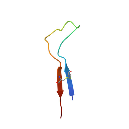
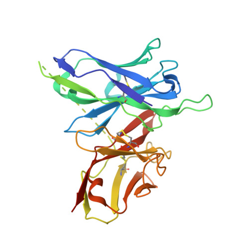
HIV envelope V3 region mimic embodies key features of a broadly neutralizing antibody lineage epitope.

Fera, D.Lee, M.S.Wiehe, K.Meyerhoff, R.R.Piai, A.Bonsignori, M.Aussedat, B.Walkowicz, W.E.Ton, T.Zhou, J.O.Danishefsky, S.Haynes, B.F.Harrison, S.C.

(2018) Nat Commun 9: 1111-1111

  • DOI: https://doi.org/10.1038/s41467-018-03565-6
  • Primary Citation Related Structures: 
    6CBJ, 6CBP

  • PubMed Abstract: 

    HIV-1 envelope (Env) mimetics are candidate components of prophylactic vaccines and potential therapeutics. Here we use a synthetic V3-glycopeptide ("Man 9 -V3") for structural studies of an HIV Env third variable loop (V3)-glycan directed, broadly neutralizing antibody (bnAb) lineage ("DH270"), to visualize the epitope on Env and to study how affinity maturation of the lineage proceeded. Unlike many previous V3 mimetics, Man 9 -V3 encompasses two key features of the V3 region recognized by V3-glycan bnAbs-the conserved GDIR motif and the N332 glycan. In our structure of an antibody fragment of a lineage member, DH270.6, in complex with the V3 glycopeptide, the conformation of the antibody-bound glycopeptide conforms closely to that of the corresponding segment in an intact HIV-1 Env trimer. An additional structure identifies roles for two critical mutations in the development of breadth. The results suggest a strategy for use of a V3 glycopeptide as a vaccine immunogen.


  • Organizational Affiliation
    • Laboratory of Molecular Medicine, Boston Children's Hospital, Harvard Medical School, Boston, MA, 02115, USA. dfera1@swarthmore.edu.

Macromolecule Content 

  • Total Structure Weight: 33.17 kDa 
  • Atom Count: 2,280 
  • Modeled Residue Count: 265 
  • Deposited Residue Count: 286 
  • Unique protein chains: 2

Macromolecules

Find similar proteins by:|  3D Structure
Entity ID: 1
MoleculeChains  Sequence LengthOrganismDetailsImage
Man9-V3 glycopeptideA [auth P]30Human immunodeficiency virus 1Mutation(s): 0 
UniProt
Find proteins for Q74448 (Human immunodeficiency virus type 1)
Explore Q74448 
Go to UniProtKB:  Q74448
Entity Groups
Sequence Clusters30% Identity50% Identity70% Identity90% Identity95% Identity100% Identity
UniProt GroupQ74448
Glycosylation
Glycosylation Sites: 1
Sequence Annotations
Expand
Reference Sequence
Find similar proteins by:|  3D Structure
Entity ID: 2
MoleculeChains  Sequence LengthOrganismDetailsImage
DH270.6 single chain variable fragmentB [auth A]256Homo sapiensMutation(s): 0 

Oligosaccharides

Help  
Entity ID: 3
MoleculeChains Length2D Diagram GlycosylationD Interactions
beta-D-mannopyranose-(1-4)-2-acetamido-2-deoxy-beta-D-glucopyranose-(1-4)-2-acetamido-2-deoxy-beta-D-glucopyranoseC [auth B]3N-Glycosylation
Glycosylation Resources
GlyTouCan: G15407YE
GlyCosmos: G15407YE
GlyGen: G15407YE
Entity ID: 4
MoleculeChains Length2D Diagram GlycosylationD Interactions
alpha-D-mannopyranose-(1-2)-alpha-D-mannopyranose-(1-2)-alpha-D-mannopyranose-(1-3)-beta-D-mannopyranose-(1-4)-2-acetamido-2-deoxy-beta-D-glucopyranose-(1-4)-2-acetamido-2-deoxy-beta-D-glucopyranoseD [auth C]6N/A
Glycosylation Resources
GlyTouCan: G01760ZU
GlyCosmos: G01760ZU
GlyGen: G01760ZU
Entity ID: 5
MoleculeChains Length2D Diagram GlycosylationD Interactions
alpha-D-mannopyranose-(1-2)-alpha-D-mannopyranose-(1-3)-alpha-D-mannopyranoseE [auth D]3N/A
Glycosylation Resources
GlyTouCan: G57891XD
GlyCosmos: G57891XD
GlyGen: G57891XD
Entity ID: 6
MoleculeChains Length2D Diagram GlycosylationD Interactions
alpha-D-mannopyranose-(1-2)-alpha-D-mannopyranoseF [auth E]2N/A
Glycosylation Resources
GlyTouCan: G53402KW
GlyCosmos: G53402KW
GlyGen: G53402KW

Biologically Interesting Molecules (External Reference) 

1 Unique

Experimental Data & Validation

Experimental Data

  • Method: X-RAY DIFFRACTION
  • Resolution: 2.17 Å
  • R-Value Free:  0.278 (Depositor), 0.276 (DCC) 
  • R-Value Work:  0.221 (Depositor), 0.231 (DCC) 
  • R-Value Observed: 0.224 (Depositor) 
Space Group: P 41 2 2
Unit Cell:
Length ( Å )Angle ( ˚ )
a = 68.005α = 90
b = 68.005β = 90
c = 128.835γ = 90
Software Package:
Software NamePurpose
PHENIXrefinement
XDSdata scaling
PHASERphasing
PDB_EXTRACTdata extraction
Cootmodel building
XDSdata reduction

Structure Validation

View Full Validation Report


Entry History 

& Funding Information

Deposition Data


Funding OrganizationLocationGrant Number
amfARUnited States109502-61-RKVA
National Institutes of Health/National Institute Of Allergy and Infectious Diseases (NIH/NIAID)United States1F32-AI-116355
National Institutes of Health/National Institute Of Allergy and Infectious Diseases (NIH/NIAID)United StatesAI100645
National Institutes of Health/National Institute of General Medical Sciences (NIH/NIGMS)United StatesT32GM007171
National Institutes of Health/National Institute Of Allergy and Infectious Diseases (NIH/NIAID)United StatesF30-AI122982
National Institutes of Health/National Institute Of Allergy and Infectious Diseases (NIH/NIAID)United StatesAI127193
National Institutes of Health/National Institute of General Medical Sciences (NIH/NIGMS)United StatesP41 GM103403

Revision History  (Full details and data files)

  • Version 1.0: 2018-02-21
    Type: Initial release
  • Version 1.1: 2018-02-28
    Changes: Author supporting evidence
  • Version 1.2: 2018-03-28
    Changes: Data collection, Database references
  • Version 1.3: 2018-04-04
    Changes: Data collection, Database references
  • Version 1.4: 2019-04-17
    Changes: Author supporting evidence, Data collection, Database references
  • Version 1.5: 2019-12-18
    Changes: Author supporting evidence
  • Version 2.0: 2020-07-29
    Type: Remediation
    Reason: Carbohydrate remediation
    Changes: Advisory, Atomic model, Data collection, Derived calculations, Structure summary
  • Version 2.1: 2023-10-04
    Changes: Data collection, Database references, Derived calculations, Refinement description, Structure summary
  • Version 2.2: 2024-11-13
    Changes: Structure summary