6C9T | pdb_00006c9t

Transcriptional repressor, CouR


Experimental Data Snapshot

  • Method: X-RAY DIFFRACTION
  • Resolution: 2.07 Å
  • R-Value Free: 
    0.334 (Depositor), 0.339 (DCC) 
  • R-Value Work: 
    0.272 (Depositor), 0.276 (DCC) 
  • R-Value Observed: 
    0.275 (Depositor) 

Starting Model: experimental
View more details

wwPDB Validation 3D Report Full Report

Validation slider image for 6C9T

This is version 1.3 of the entry. See complete history

Literature

Structural basis of transcriptional regulation by CouR, a repressor of coumarate catabolism, inRhodopseudomonas palustris.

Cogan, D.P.Baraquet, C.Harwood, C.S.Nair, S.K.

(2018) J Biological Chem 293: 11727-11735

  • DOI: https://doi.org/10.1074/jbc.RA118.003561
  • Primary Citation Related Structures: 
    6C28, 6C2S, 6C9T

  • PubMed Abstract: 

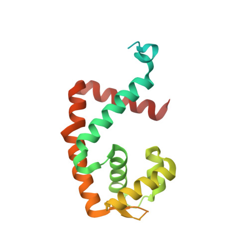
    The MarR family transcriptional regulator CouR, from the soil bacterium Rhodopseudomonas palustris CGA009, has recently been shown to negatively regulate a p -coumarate catabolic operon. Unlike most characterized MarR repressors that respond to small metabolites at concentrations in the millimolar range, repression by CouR is alleviated by the 800-Da ligand p -coumaroyl-CoA with high affinity and specificity. Here we report the crystal structures of ligand-free CouR as well as the complex with p -coumaroyl-CoA, each to 2.1-Å resolution, and the 2.85-Å resolution cocrystal structure of CouR bound to an oligonucleotide bearing the cognate DNA operator sequence. In combination with binding experiments that uncover specific residues important for ligand and DNA recognition, these structures provide glimpses of a MarR family repressor in all possible states, providing an understanding of the molecular basis of DNA binding and the conformation alterations that accompany ligand-induced dissociation for activation of the operon.


  • Organizational Affiliation
    • From the Department of Biochemistry.

Macromolecule Content 

  • Total Structure Weight: 40.51 kDa 
  • Atom Count: 2,410 
  • Modeled Residue Count: 269 
  • Deposited Residue Count: 372 
  • Unique protein chains: 1

Macromolecules

Find similar proteins by:|  3D Structure
Entity ID: 1
MoleculeChains  Sequence LengthOrganismDetailsImage
CouR
A, B
186Rhodopseudomonas palustris CGA009Mutation(s): 0 
Gene Names: RPA1794
UniProt
Find proteins for Q6N8V9 (Rhodopseudomonas palustris (strain ATCC BAA-98 / CGA009))
Explore Q6N8V9 
Go to UniProtKB:  Q6N8V9
Entity Groups
Sequence Clusters30% Identity50% Identity70% Identity90% Identity95% Identity100% Identity
UniProt GroupQ6N8V9
Sequence Annotations
Expand
Reference Sequence

Experimental Data & Validation

Experimental Data

  • Method: X-RAY DIFFRACTION
  • Resolution: 2.07 Å
  • R-Value Free:  0.334 (Depositor), 0.339 (DCC) 
  • R-Value Work:  0.272 (Depositor), 0.276 (DCC) 
  • R-Value Observed: 0.275 (Depositor) 
Space Group: P 41
Unit Cell:
Length ( Å )Angle ( ˚ )
a = 59.008α = 90
b = 59.008β = 90
c = 108.402γ = 90
Software Package:
Software NamePurpose
REFMACrefinement
XDSdata reduction
XDSdata scaling
PHASERphasing

Structure Validation

View Full Validation Report


Entry History 

Deposition Data

Revision History  (Full details and data files)

  • Version 1.0: 2018-05-30
    Type: Initial release
  • Version 1.1: 2018-06-13
    Changes: Data collection, Database references
  • Version 1.2: 2018-08-08
    Changes: Data collection, Database references
  • Version 1.3: 2023-10-04
    Changes: Data collection, Database references, Refinement description