6A72 | pdb_00006a72

Copper transporter protein


Experimental Data Snapshot

  • Method: X-RAY DIFFRACTION
  • Resolution: 2.10 Å
  • R-Value Free: 
    0.211 (Depositor), 0.209 (DCC) 
  • R-Value Work: 
    0.174 (Depositor), 0.182 (DCC) 
  • R-Value Observed: 
    0.176 (Depositor) 

Starting Model: experimental
View more details

wwPDB Validation 3D Report Full Report

Validation slider image for 6A72

This is version 1.2 of the entry. See complete history

Literature

Tetrathiomolybdate induces dimerization of the metal-binding domain of ATPase and inhibits platination of the protein.

Fang, T.Chen, W.Sheng, Y.Yuan, S.Tang, Q.Li, G.Huang, G.Su, J.Zhang, X.Zang, J.Liu, Y.

(2019) Nat Commun 10: 186-186

  • DOI: https://doi.org/10.1038/s41467-018-08102-z
  • Primary Citation Related Structures: 
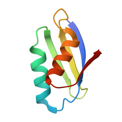
    6A71, 6A72

  • PubMed Abstract: 

    Tetrathiomolybdate (TM) is used in the clinic for the treatment of Wilson's disease by targeting the cellular copper efflux protein ATP7B (WLN). Interestingly, both TM and WLN are associated with the efficacy of cisplatin, a widely used anticancer drug. Herein, we show that TM induces dimerization of the metal-binding domain of ATP7B (WLN4) through a unique sulfur-bridged Mo 2 S 6 O 2 cluster. TM expels copper ions from Cu-WLN4 and forms a copper-free dimer. The binding of Mo to cysteine residues of WLN4 inhibits platination of the protein. Reaction with multi-domain proteins indicates that TM can also connect two domains in the same molecule, forming Mo-bridged intramolecular crosslinks. These results provide structural and chemical insight into the mechanism of action of TM against ATPase, and reveal the molecular mechanism by which TM attenuates the cisplatin resistance mediated by copper efflux proteins.


  • Organizational Affiliation
    • CAS Key Laboratory of Soft Matter Chemistry, Department of Chemistry, University of Science and Technology of China, Hefei, Anhui, 230026, China.

Macromolecule Content 

  • Total Structure Weight: 15.14 kDa 
  • Atom Count: 1,118 
  • Modeled Residue Count: 143 
  • Deposited Residue Count: 144 
  • Unique protein chains: 1

Macromolecules

Find similar proteins by:|  3D Structure
Entity ID: 1
MoleculeChains  Sequence LengthOrganismDetailsImage
ATP7B protein
A, B
72Homo sapiensMutation(s): 0 
Gene Names: ATP7B
EC: 7.2.2.8
UniProt & NIH Common Fund Data Resources
Find proteins for P35670 (Homo sapiens)
Explore P35670 
Go to UniProtKB:  P35670
PHAROS:  P35670
GTEx:  ENSG00000123191 
Entity Groups
Sequence Clusters30% Identity50% Identity70% Identity90% Identity95% Identity100% Identity
UniProt GroupP35670
Sequence Annotations
Expand
Reference Sequence

Small Molecules

Experimental Data & Validation

Experimental Data

  • Method: X-RAY DIFFRACTION
  • Resolution: 2.10 Å
  • R-Value Free:  0.211 (Depositor), 0.209 (DCC) 
  • R-Value Work:  0.174 (Depositor), 0.182 (DCC) 
  • R-Value Observed: 0.176 (Depositor) 
Space Group: C 2 2 21
Unit Cell:
Length ( Å )Angle ( ˚ )
a = 46.114α = 90
b = 87.582β = 90
c = 77.301γ = 90
Software Package:
Software NamePurpose
REFMACrefinement
HKL-2000data reduction
HKL-2000data scaling
PHASERphasing

Structure Validation

View Full Validation Report


Entry History 

Deposition Data

  • Released Date: 2019-04-03 
  • Deposition Author(s): Chen, W.B.

Revision History  (Full details and data files)

  • Version 1.0: 2019-04-03
    Type: Initial release
  • Version 1.1: 2019-08-28
    Changes: Data collection, Derived calculations
  • Version 1.2: 2023-11-22
    Changes: Data collection, Database references, Derived calculations, Refinement description