6A57 | pdb_00006a57

Structure of histone demethylase REF6 complexed with DNA


Experimental Data Snapshot

  • Method: X-RAY DIFFRACTION
  • Resolution: 2.70 Å
  • R-Value Free: 
    0.257 (Depositor), 0.258 (DCC) 
  • R-Value Work: 
    0.221 (Depositor), 0.225 (DCC) 
  • R-Value Observed: 
    0.223 (Depositor) 

wwPDB Validation 3D Report Full Report

Validation slider image for 6A57

This is version 1.2 of the entry. See complete history

Literature

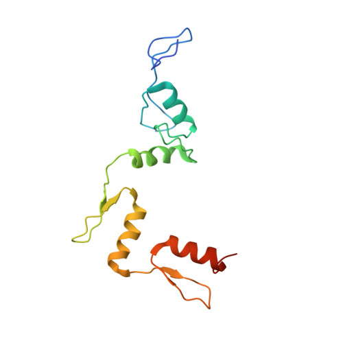
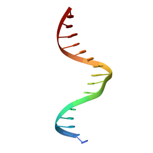
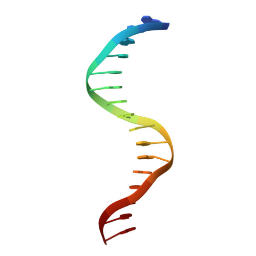
Crystal structures of REF6 and its complex with DNA reveal diverse recognition mechanisms.

Tian, Z.Li, X.Li, M.Wu, W.Zhang, M.Tang, C.Li, Z.Liu, Y.Chen, Z.Yang, M.Ma, L.Caba, C.Tong, Y.Lam, H.M.Dai, S.Chen, Z.

(2020) Cell Discov 6: 17-17

  • DOI: https://doi.org/10.1038/s41421-020-0150-6
  • Primary Citation Related Structures: 
    6A57, 6A58, 6A59

  • PubMed Abstract: 

    Relative of Early Flowing 6 (REF6) is a DNA-sequence-specific H3K27me3/2 demethylase that contains four zinc finger (ZnF) domains and targets several thousand genes in Arabidopsis thaliana . The ZnF domains are essential for binding target genes, but the structural basis remains unclear. Here, we determined crystal structures of the ZnF domains and REF6-DNA complex, revealing a unique REF6-family-specific half-cross-braced ZnF (RCZ) domain and two C2H2-type ZnFs. DNA-binding induces a profound conformational change in the hinge region of REF6. Each REF6 recognizes six bases and DNA methylation reduces the binding affinity. Both the acidic region and basic region are important for the self-association of REF6. The REF6 DNA-binding affinity is determined by the sequence-dependent conformations of DNA and also the cooperativity in different target motifs. The conformational plasticity enables REF6 to function as a global transcriptional regulator that directly binds to many diverse genes, revealing the structural basis for the epigenetic modification recognition.


  • Organizational Affiliation
    • 1State Key Laboratory of Agrobiotechnology and Beijing Advanced Innovation Center for Food Nutrition and Human Health, College of Biological Sciences, China Agricultural University, 100193 Beijing, China.

Macromolecule Content 

  • Total Structure Weight: 26.76 kDa 
  • Atom Count: 1,746 
  • Modeled Residue Count: 163 
  • Deposited Residue Count: 172 
  • Unique protein chains: 1
  • Unique nucleic acid chains: 2

Macromolecules


Find similar proteins by:|  3D Structure
Entity ID: 1
MoleculeChains  Sequence LengthOrganismDetailsImage
Lysine-specific demethylase REF6140Arabidopsis thalianaMutation(s): 0 
Gene Names: REF6JMJ12PKDM9AAt3g48430T29H11_50
EC: 1.14.11
UniProt
Find proteins for Q9STM3 (Arabidopsis thaliana)
Explore Q9STM3 
Go to UniProtKB:  Q9STM3
Entity Groups
Sequence Clusters30% Identity50% Identity70% Identity90% Identity95% Identity100% Identity
UniProt GroupQ9STM3
Sequence Annotations
Expand
Reference Sequence
Find similar nucleic acids by:  Sequence
Entity ID: 2
MoleculeChains LengthOrganismImage
DNA (5'-D(*CP*TP*TP*TP*CP*TP*CP*TP*GP*TP*TP*TP*TP*GP*TP*C)-3')16Arabidopsis thaliana
Sequence Annotations
Expand
Reference Sequence
Find similar nucleic acids by:  Sequence
Entity ID: 3
MoleculeChains LengthOrganismImage
DNA (5'-D(*GP*GP*AP*CP*AP*AP*AP*AP*CP*AP*GP*AP*GP*AP*AP*A)-3')16Arabidopsis thaliana
Sequence Annotations
Expand
Reference Sequence

Experimental Data & Validation

Experimental Data

  • Method: X-RAY DIFFRACTION
  • Resolution: 2.70 Å
  • R-Value Free:  0.257 (Depositor), 0.258 (DCC) 
  • R-Value Work:  0.221 (Depositor), 0.225 (DCC) 
  • R-Value Observed: 0.223 (Depositor) 
Space Group: P 32 2 1
Unit Cell:
Length ( Å )Angle ( ˚ )
a = 96.087α = 90
b = 96.087β = 90
c = 73.905γ = 120
Software Package:
Software NamePurpose
REFMACrefinement
HKL-2000data reduction
HKL-2000data scaling
HKL2Mapphasing

Structure Validation

View Full Validation Report


Entry History 

& Funding Information

Deposition Data


Funding OrganizationLocationGrant Number
National Basic Research Program of China (973 Program)China2016YFC1200400

Revision History  (Full details and data files)

  • Version 1.0: 2019-06-26
    Type: Initial release
  • Version 1.1: 2020-07-08
    Changes: Author supporting evidence, Database references
  • Version 1.2: 2024-03-27
    Changes: Data collection, Database references