


Crystal structure of the capping enzyme P5 from Rice Dwarf Virus
Nakamichi, Y., Higashiura, A., Narita, H., Hagiwara, K., Uehara-Ichiki, T., Omura, T., Nakagawa, A.To be published.
Experimental Data Snapshot
Starting Model: experimental
View more details
Entity ID: 1 | |||||
|---|---|---|---|---|---|
| Molecule | Chains | Sequence Length | Organism | Details | Image |
| mRNA capping enzyme P5 | 804 | Rice dwarf virus (isolate O) | Mutation(s): 0 EC: 2.7.7.50 | 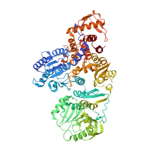 | |
UniProt | |||||
Entity Groups | |||||
| Sequence Clusters | 30% Identity50% Identity70% Identity90% Identity95% Identity100% Identity | ||||
| UniProt Group | P14583 | ||||
Sequence AnnotationsExpand | |||||
Reference Sequence | |||||
| Ligands 3 Unique | |||||
|---|---|---|---|---|---|
| ID | Chains | Name / Formula / InChI Key | 2D Diagram | 3D Interactions | |
| SAM Download:Ideal Coordinates CCD File | E [auth A], F [auth A], K [auth B], P [auth C], Q [auth C] | S-ADENOSYLMETHIONINE C15 H22 N6 O5 S MEFKEPWMEQBLKI-FCKMPRQPSA-N |  | ||
| FLC Download:Ideal Coordinates CCD File | J [auth A], O [auth B], U [auth D] | CITRATE ANION C6 H5 O7 KRKNYBCHXYNGOX-UHFFFAOYSA-K |  | ||
| EDO Download:Ideal Coordinates CCD File | G [auth A] H [auth A] I [auth A] L [auth B] M [auth B] | 1,2-ETHANEDIOL C2 H6 O2 LYCAIKOWRPUZTN-UHFFFAOYSA-N |  | ||
| Length ( Å ) | Angle ( ˚ ) |
|---|---|
| a = 60.182 | α = 73.8 |
| b = 122.778 | β = 87.14 |
| c = 129.257 | γ = 86.08 |
| Software Name | Purpose |
|---|---|
| PHENIX | refinement |
| HKL-2000 | data reduction |
| HKL-2000 | data scaling |
| MOLREP | phasing |
| Funding Organization | Location | Grant Number |
|---|---|---|
| JSPS | Japan | JP25251009 |


