5IQZ | pdb_00005iqz

Crystal structure of N-terminal domain of Human SIRT7


Experimental Data Snapshot

  • Method: X-RAY DIFFRACTION
  • Resolution: 2.33 Å
  • R-Value Free: 
    0.237 (Depositor), 0.235 (DCC) 
  • R-Value Work: 
    0.183 (Depositor), 0.186 (DCC) 
  • R-Value Observed: 
    0.186 (Depositor) 

Starting Model: experimental
View more details

wwPDB Validation 3D Report Full Report

Validation slider image for 5IQZ

This is version 2.1 of the entry. See complete history

Literature

Crystal structure of the N-terminal domain of human SIRT7 reveals a three-helical domain architecture

Priyanka, A.Solanki, V.Parkesh, R.Thakur, K.G.

(2016) Proteins 84: 1558-1563

  • DOI: https://doi.org/10.1002/prot.25085
  • Primary Citation Related Structures: 
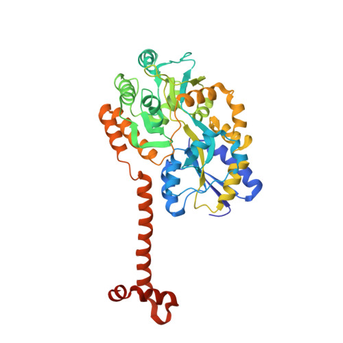
    5IQZ

  • PubMed Abstract: 

    Human SIRT7 is an NAD(+) dependent deacetylase, which belongs to sirtuin family of proteins. SIRT7, like other sirtuins has conserved catalytic domain and is flanked by N- and C-terminal domains reported to play vital functional roles. Here, we report the crystal structure of the N-terminal domain of human SIRT7 (SIRT7(NTD) ) at 2.3 Å resolution as MBP-SIRT7(NTD) fusion protein. SIRT7(NTD) adopts three-helical domain architecture and comparative structural analyses suggest similarities to some DNA binding motifs and transcription regulators. We also report here the importance of N- and C-terminal domains in soluble expression of SIRT7. Proteins 2016; 84:1558-1563. © 2016 Wiley Periodicals, Inc.


  • Organizational Affiliation
    • Structural Biology Laboratory, G. N. Ramachandran Protein Centre, CSIR-Institute of Microbial Technology, Chandigarh, 160036, India.

Macromolecule Content 

  • Total Structure Weight: 49.51 kDa 
  • Atom Count: 3,558 
  • Modeled Residue Count: 426 
  • Deposited Residue Count: 443 
  • Unique protein chains: 1

Macromolecules

Find similar proteins by:|  3D Structure
Entity ID: 1
MoleculeChains  Sequence LengthOrganismDetailsImage
SIRT7 protein443Homo sapiensMutation(s): 0 
Gene Names: sirt7
EC: 2.3.1 (UniProt), 2.3.1.286 (UniProt)
UniProt & NIH Common Fund Data Resources
Find proteins for P0AEX9 (Escherichia coli (strain K12))
Explore P0AEX9 
Go to UniProtKB:  P0AEX9
Find proteins for Q9NRC8 (Homo sapiens)
Explore Q9NRC8 
Go to UniProtKB:  Q9NRC8
PHAROS:  Q9NRC8
GTEx:  ENSG00000187531 
Entity Groups
Sequence Clusters30% Identity50% Identity70% Identity90% Identity95% Identity100% Identity
UniProt GroupsQ9NRC8P0AEX9
Sequence Annotations
Expand
Reference Sequence

Oligosaccharides

Help  
Entity ID: 2
MoleculeChains Length2D Diagram GlycosylationD Interactions
alpha-D-glucopyranose-(1-4)-alpha-D-glucopyranose
B
2N/A
Glycosylation Resources
GlyTouCan: G07411ON
GlyCosmos: G07411ON

Biologically Interesting Molecules (External Reference) 

1 Unique

Experimental Data & Validation

Experimental Data

  • Method: X-RAY DIFFRACTION
  • Resolution: 2.33 Å
  • R-Value Free:  0.237 (Depositor), 0.235 (DCC) 
  • R-Value Work:  0.183 (Depositor), 0.186 (DCC) 
  • R-Value Observed: 0.186 (Depositor) 
Space Group: P 1 21 1
Unit Cell:
Length ( Å )Angle ( ˚ )
a = 60.49α = 90
b = 49.33β = 101.2
c = 65.68γ = 90
Software Package:
Software NamePurpose
PHENIXrefinement
MOSFLMdata processing
SCALAdata scaling
PHASERphasing
PDB_EXTRACTdata extraction
Cootmodel building
iMOSFLMdata reduction

Structure Validation

View Full Validation Report


Entry History 

Deposition Data

Revision History  (Full details and data files)

  • Version 1.0: 2016-07-27
    Type: Initial release
  • Version 1.1: 2016-09-28
    Changes: Database references
  • Version 2.0: 2020-07-29
    Type: Remediation
    Reason: Carbohydrate remediation
    Changes: Atomic model, Data collection, Database references, Derived calculations, Non-polymer description, Structure summary
  • Version 2.1: 2023-11-08
    Changes: Data collection, Database references, Refinement description, Structure summary