5A6C | pdb_00005a6c

Concomitant binding of Afadin to LGN and F-actin directs planar spindle orientation


Experimental Data Snapshot

  • Method: X-RAY DIFFRACTION
  • Resolution: 2.90 Å
  • R-Value Free: 
    0.252 (Depositor), 0.250 (DCC) 
  • R-Value Work: 
    0.206 (Depositor), 0.208 (DCC) 
  • R-Value Observed: 
    0.208 (Depositor) 

Starting Model: experimental
View more details

wwPDB Validation 3D Report Full Report

Validation slider image for 5A6C

This is version 1.4 of the entry. See complete history

Literature

Concomitant Binding of Afadin to Lgn and F-Actin Directs Planar Spindle Orientation.

Carminati, M.Gallini, S.Pirovano, L.Alfieri, A.Bisi, S.Mapelli, M.

(2016) Nat Struct Mol Biol 23: 155

  • DOI: https://doi.org/10.1038/nsmb.3152
  • Primary Citation Related Structures: 
    5A6C

  • PubMed Abstract: 

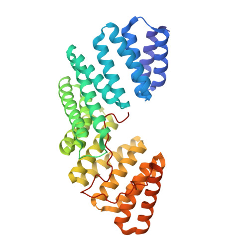
    Polarized epithelia form by oriented cell divisions in which the mitotic spindle aligns parallel to the epithelial plane. To orient the mitotic spindle, cortical cues trigger the recruitment of NuMA-dynein-based motors, which pull on astral microtubules via the protein LGN. We demonstrate that the junctional protein Afadin is required for spindle orientation and correct epithelial morphogenesis of Caco-2 cysts. Molecularly, Afadin binds directly and concomitantly to F-actin and to LGN. We determined the crystallographic structure of human Afadin in complex with LGN and show that it resembles the LGN-NuMA complex. In mitosis, Afadin is necessary for cortical accumulation of LGN and NuMA above the spindle poles, in an F-actin-dependent manner. Collectively, our results depict Afadin as a molecular hub governing the enrichment of LGN and NuMA at the cortex. To our knowledge, Afadin is the first-described mechanical anchor between dynein and cortical F-actin.


  • Organizational Affiliation
    • Department of Experimental Oncology, European Institute of Oncology, Milan, Italy.

Macromolecule Content 

  • Total Structure Weight: 83.29 kDa 
  • Atom Count: 5,747 
  • Modeled Residue Count: 732 
  • Deposited Residue Count: 748 
  • Unique protein chains: 1

Macromolecules

Find similar proteins by:|  3D Structure
Entity ID: 1
MoleculeChains  Sequence LengthOrganismDetailsImage
G-PROTEIN-SIGNALING MODULATOR 2, AFADIN
A, B
374Homo sapiensMutation(s): 0 
Gene Names: GPSM2LGNMLLT4AF6
UniProt & NIH Common Fund Data Resources
Find proteins for P55196 (Homo sapiens)
Explore P55196 
Go to UniProtKB:  P55196
PHAROS:  P55196
GTEx:  ENSG00000130396 
Find proteins for P81274 (Homo sapiens)
Explore P81274 
Go to UniProtKB:  P81274
PHAROS:  P81274
GTEx:  ENSG00000121957 
Entity Groups
Sequence Clusters30% Identity50% Identity70% Identity90% Identity95% Identity100% Identity
UniProt GroupsP55196P81274
Sequence Annotations
Expand
Reference Sequence

Experimental Data & Validation

Experimental Data

  • Method: X-RAY DIFFRACTION
  • Resolution: 2.90 Å
  • R-Value Free:  0.252 (Depositor), 0.250 (DCC) 
  • R-Value Work:  0.206 (Depositor), 0.208 (DCC) 
  • R-Value Observed: 0.208 (Depositor) 
Space Group: P 21 3
Unit Cell:
Length ( Å )Angle ( ˚ )
a = 169.88α = 90
b = 169.88β = 90
c = 169.88γ = 90
Software Package:
Software NamePurpose
PHENIXrefinement
XDSdata reduction
SCALAdata scaling
PHASERphasing

Structure Validation

View Full Validation Report


Entry History 

Deposition Data

Revision History  (Full details and data files)

  • Version 1.0: 2015-12-30
    Type: Initial release
  • Version 1.1: 2016-01-20
    Changes: Database references
  • Version 1.2: 2016-01-27
    Changes: Structure summary
  • Version 1.3: 2016-03-02
    Changes: Database references
  • Version 1.4: 2024-01-10
    Changes: Data collection, Database references, Derived calculations, Other, Refinement description