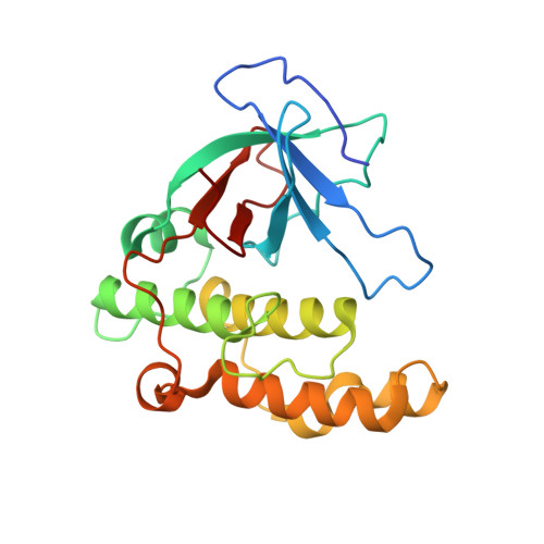
5A0L | pdb_00005a0l

N-terminal thioester domain of fibronectin-binding protein SfbI from Streptococcus pyogenes


Experimental Data Snapshot

  • Method: X-RAY DIFFRACTION
  • Resolution: 1.35 Å
  • R-Value Free: 
    0.153 (Depositor), 0.165 (DCC) 
  • R-Value Work: 
    0.131 (Depositor), 0.141 (DCC) 
  • R-Value Observed: 
    0.132 (Depositor) 

wwPDB Validation 3D Report Full Report

Validation slider image for 5A0L

This is version 1.4 of the entry. See complete history. 

Literature

An internal thioester in a pathogen surface protein mediates covalent host binding.

Walden, M., Edwards, J.M., Dziewulska, A.M., Bergmann, R., Saalbach, G., Kan, S.Y., Miller, O.K., Weckener, M., Jackson, R.J., Shirran, S.L., Botting, C.H., Florence, G.J., Rohde, M., Banfield, M.J., Schwarz-Linek, U.

(2015) Elife 4

  • DOI: https://doi.org/10.7554/eLife.06638
  • Primary Citation Related Structures: 
    5A0D, 5A0G, 5A0L, 5A0N

  • PubMed Abstract: 

    To cause disease and persist in a host, pathogenic and commensal microbes must adhere to tissues. Colonization and infection depend on specific molecular interactions at the host-microbe interface that involve microbial surface proteins, or adhesins. To date, adhesins are only known to bind to host receptors non-covalently. Here we show that the streptococcal surface protein SfbI mediates covalent interaction with the host protein fibrinogen using an unusual internal thioester bond as a 'chemical harpoon'. This cross-linking reaction allows bacterial attachment to fibrin and SfbI binding to human cells in a model of inflammation. Thioester-containing domains are unexpectedly prevalent in Gram-positive bacteria, including many clinically relevant pathogens. Our findings support bacterial-encoded covalent binding as a new molecular principle in host-microbe interactions. This represents an as yet unexploited target to treat bacterial infection and may also offer novel opportunities for engineering beneficial interactions.


  • Organizational Affiliation: 
    • Department of Biological Chemistry, John Innes Centre, Norwich, United Kingdom.

Macromolecule Content 

  • Total Structure Weight: 48.81 kDa 
  • Atom Count: 3,859 
  • Modeled Residue Count: 410 
  • Deposited Residue Count: 420 
  • Unique protein chains: 1

Macromolecules

Find similar proteins by:|  3D Structure
Entity ID: 1
MoleculeChains  Sequence LengthOrganismDetailsImage
FIBRONECTIN-BINDING PROTEIN
A, B
210Streptococcus pyogenes MGAS2096Mutation(s): 0 
UniProt
Find proteins for A0A0M3KL43 (Streptococcus pyogenes serotype M12 (strain MGAS2096))
Explore A0A0M3KL43 
Go to UniProtKB:  A0A0M3KL43
Entity Groups
Sequence Clusters30% Identity50% Identity70% Identity90% Identity95% Identity100% Identity
UniProt GroupA0A0M3KL43
Sequence Annotations
Expand
Reference Sequence

Experimental Data & Validation

Experimental Data

  • Method: X-RAY DIFFRACTION
  • Resolution: 1.35 Å
  • R-Value Free:  0.153 (Depositor), 0.165 (DCC) 
  • R-Value Work:  0.131 (Depositor), 0.141 (DCC) 
  • R-Value Observed: 0.132 (Depositor) 
Space Group: I 41
Unit Cell:
Length ( Å )Angle ( ˚ )
a = 165.12α = 90
b = 165.12β = 90
c = 42.52γ = 90
Software Package:
Software NamePurpose
REFMACrefinement
xia2data reduction
xia2data scaling
PHENIXphasing

Structure Validation

View Full Validation Report


Entry History 

Deposition Data

Revision History  (Full details and data files)

  • Version 1.0: 2015-06-03
    Type: Initial release
  • Version 1.1: 2015-06-10
    Changes: Database references
  • Version 1.2: 2017-03-22
    Changes: Database references
  • Version 1.3: 2019-10-30
    Changes: Advisory, Data collection, Derived calculations, Other
  • Version 1.4: 2024-05-08
    Changes: Data collection, Database references, Derived calculations