5A0N | pdb_00005a0n

N-terminal thioester domain of protein F2 like fibronectin-binding protein from Streptococcus pneumoniae


Experimental Data Snapshot

  • Method: X-RAY DIFFRACTION
  • Resolution: 1.30 Å
  • R-Value Free: 
    0.151 (Depositor), 0.165 (DCC) 
  • R-Value Work: 
    0.119 (Depositor), 0.135 (DCC) 
  • R-Value Observed: 
    0.121 (Depositor) 

wwPDB Validation 3D Report Full Report

Validation slider image for 5A0N

This is version 1.4 of the entry. See complete history

Literature

An internal thioester in a pathogen surface protein mediates covalent host binding.

Walden, M.Edwards, J.M.Dziewulska, A.M.Bergmann, R.Saalbach, G.Kan, S.Y.Miller, O.K.Weckener, M.Jackson, R.J.Shirran, S.L.Botting, C.H.Florence, G.J.Rohde, M.Banfield, M.J.Schwarz-Linek, U.

(2015) Elife 4

  • DOI: https://doi.org/10.7554/eLife.06638
  • Primary Citation Related Structures: 
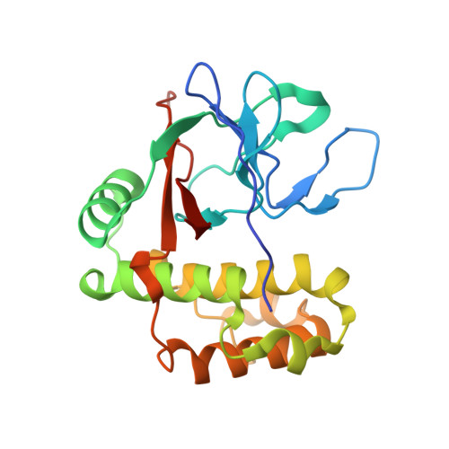
    5A0D, 5A0G, 5A0L, 5A0N

  • PubMed Abstract: 

    To cause disease and persist in a host, pathogenic and commensal microbes must adhere to tissues. Colonization and infection depend on specific molecular interactions at the host-microbe interface that involve microbial surface proteins, or adhesins. To date, adhesins are only known to bind to host receptors non-covalently. Here we show that the streptococcal surface protein SfbI mediates covalent interaction with the host protein fibrinogen using an unusual internal thioester bond as a 'chemical harpoon'. This cross-linking reaction allows bacterial attachment to fibrin and SfbI binding to human cells in a model of inflammation. Thioester-containing domains are unexpectedly prevalent in Gram-positive bacteria, including many clinically relevant pathogens. Our findings support bacterial-encoded covalent binding as a new molecular principle in host-microbe interactions. This represents an as yet unexploited target to treat bacterial infection and may also offer novel opportunities for engineering beneficial interactions.


  • Organizational Affiliation
    • Department of Biological Chemistry, John Innes Centre, Norwich, United Kingdom.

Macromolecule Content 

  • Total Structure Weight: 26.1 kDa 
  • Atom Count: 2,100 
  • Modeled Residue Count: 214 
  • Deposited Residue Count: 220 
  • Unique protein chains: 1

Macromolecules

Find similar proteins by:|  3D Structure
Entity ID: 1
MoleculeChains  Sequence LengthOrganismDetailsImage
PROTEIN F2 LIKE FIBRONECTIN-BINDING PROTEIN220Streptococcus pneumoniae SP14-BS69Mutation(s): 0 

Small Molecules

Experimental Data & Validation

Experimental Data

  • Method: X-RAY DIFFRACTION
  • Resolution: 1.30 Å
  • R-Value Free:  0.151 (Depositor), 0.165 (DCC) 
  • R-Value Work:  0.119 (Depositor), 0.135 (DCC) 
  • R-Value Observed: 0.121 (Depositor) 
Space Group: P 41 21 2
Unit Cell:
Length ( Å )Angle ( ˚ )
a = 59.86α = 90
b = 59.86β = 90
c = 121.66γ = 90
Software Package:
Software NamePurpose
REFMACrefinement
MOSFLMdata reduction
Aimlessdata scaling
PHENIXphasing

Structure Validation

View Full Validation Report


Entry History 

Deposition Data

Revision History  (Full details and data files)

  • Version 1.0: 2015-06-03
    Type: Initial release
  • Version 1.1: 2015-06-10
    Changes: Database references
  • Version 1.2: 2017-03-22
    Changes: Database references
  • Version 1.3: 2019-10-30
    Changes: Advisory, Data collection, Derived calculations, Other
  • Version 1.4: 2024-05-08
    Changes: Data collection, Database references, Derived calculations