5XN1 | pdb_00005xn1

HIV-1 reverse transcriptase Q151M:DNA:entecavir-triphosphate ternary complex


Experimental Data Snapshot

  • Method: X-RAY DIFFRACTION
  • Resolution: 2.45 Å
  • R-Value Free: 
    0.227 (Depositor), 0.230 (DCC) 
  • R-Value Work: 
    0.188 (Depositor), 0.191 (DCC) 
  • R-Value Observed: 
    0.190 (Depositor) 

Starting Model: experimental
View more details

wwPDB Validation 3D Report Full Report

Validation slider image for 5XN1

Ligand Structure Quality Assessment 


This is version 2.1 of the entry. See complete history

Literature

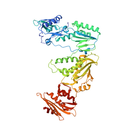
HIV-1 with HBV-associated Q151M substitution in RT becomes highly susceptible to entecavir: structural insights into HBV-RT inhibition by entecavir.

Yasutake, Y.Hattori, S.I.Hayashi, H.Matsuda, K.Tamura, N.Kohgo, S.Maeda, K.Mitsuya, H.

(2018) Sci Rep 8: 1624-1624

  • DOI: https://doi.org/10.1038/s41598-018-19602-9
  • Primary Citation Related Structures: 
    5XN0, 5XN1, 5XN2

  • PubMed Abstract: 

    Hepatitis B virus (HBV) reverse transcriptase (RT) is essential for viral replication and is an important drug target. Nonetheless, the notorious insolubility of HBV RT has hindered experimental structural studies and structure-based drug design. Here, we demonstrate that a Q151M substitution alone at the nucleotide-binding site (N-site) of human immunodeficiency virus type-1 (HIV-1) RT renders HIV-1 highly sensitive to entecavir (ETV), a potent nucleoside analogue RT inhibitor (NRTI) against HBV. The results suggest that Met151 forms a transient hydrophobic interaction with the cyclopentyl methylene of ETV, a characteristic hydrophobic moiety of ETV. We thus solved the crystal structures of HIV-1 RT Q151M :DNA complex with bound dGTP or ETV-triphosphate (ETV-TP). The structures revealed that ETV-TP is accommodated at the N-site slightly apart from the ribose ring of the 3'-end nucleotide, compared to the position of bound dGTP and previously reported NRTI/dNTP. In addition, the protruding methylene group of bound ETV-TP directly pushes the side-chain of Met184 backward. Met184 is a key residue that confers ETV resistance upon substitution with smaller Ile/Val. These results provide novel insights into NRTI binding to the N-site and further provide important clues for the development of novel anti-HBV/HIV-1 RT inhibitors to overcome critical drug resistance.


  • Organizational Affiliation
    • Bioproduction Research Institute, National Institute of Advanced Industrial Science and Technology (AIST), Sapporo, 062-8517, Japan. y-yasutake@aist.go.jp.

Macromolecule Content 

  • Total Structure Weight: 257.12 kDa 
  • Atom Count: 17,562 
  • Modeled Residue Count: 1,989 
  • Deposited Residue Count: 2,078 
  • Unique protein chains: 2
  • Unique nucleic acid chains: 1

Macromolecules


Find similar proteins by:|  3D Structure
Entity ID: 1
MoleculeChains  Sequence LengthOrganismDetailsImage
Pol proteinA,
D [auth C]
557Human immunodeficiency virus 1Mutation(s): 3 
Gene Names: pol
UniProt
Find proteins for P12497 (Human immunodeficiency virus type 1 group M subtype B (isolate NY5))
Explore P12497 
Go to UniProtKB:  P12497
Entity Groups
Sequence Clusters30% Identity50% Identity70% Identity90% Identity95% Identity100% Identity
UniProt GroupP12497
Sequence Annotations
Expand
Reference Sequence
Find similar proteins by:|  3D Structure
Entity ID: 2
MoleculeChains  Sequence LengthOrganismDetailsImage
Pol proteinB,
E [auth D]
444Human immunodeficiency virus 1Mutation(s): 2 
Gene Names: pol
UniProt
Find proteins for P12497 (Human immunodeficiency virus type 1 group M subtype B (isolate NY5))
Explore P12497 
Go to UniProtKB:  P12497
Entity Groups
Sequence Clusters30% Identity50% Identity70% Identity90% Identity95% Identity100% Identity
UniProt GroupP12497
Sequence Annotations
Expand
Reference Sequence
Find similar nucleic acids by:  (by identity cutoff) 
Entity ID: 3
MoleculeChains LengthOrganismImage
38-MER DNA aptamerC [auth E],
F
38synthetic construct
Sequence Annotations
Expand
Reference Sequence

Oligosaccharides

Help  
Entity ID: 4
MoleculeChains Length2D Diagram GlycosylationD Interactions
beta-D-fructofuranose-(2-1)-alpha-D-glucopyranose
G
2N/A
Glycosylation Resources
GlyTouCan: G05551OP
GlyCosmos: G05551OP

Small Molecules

Ligands 3 Unique
IDChains Name / Formula / InChI Key2D Diagram3D Interactions
ET9

Query on ET9


Download:Ideal Coordinates CCD File
H [auth A],
L [auth C]
[[(1R,3S,5S)-3-(2-azanyl-6-oxidanylidene-3H-purin-9-yl)-2-methylidene-5-oxidanyl-cyclopentyl]methoxy-oxidanyl-phosphory l] phosphono hydrogen phosphate
C12 H18 N5 O12 P3
YMBBDUCQYPKKJK-FXQIFTODSA-N
GOL

Query on GOL


Download:Ideal Coordinates CCD File
J [auth B],
K [auth B],
N [auth D],
O [auth D],
P [auth F]
GLYCEROL
C3 H8 O3
PEDCQBHIVMGVHV-UHFFFAOYSA-N
MG

Query on MG


Download:Ideal Coordinates CCD File
I [auth A],
M [auth C]
MAGNESIUM ION
Mg
JLVVSXFLKOJNIY-UHFFFAOYSA-N

Biologically Interesting Molecules (External Reference) 

1 Unique

Experimental Data & Validation

Experimental Data

  • Method: X-RAY DIFFRACTION
  • Resolution: 2.45 Å
  • R-Value Free:  0.227 (Depositor), 0.230 (DCC) 
  • R-Value Work:  0.188 (Depositor), 0.191 (DCC) 
  • R-Value Observed: 0.190 (Depositor) 
Space Group: H 3
Unit Cell:
Length ( Å )Angle ( ˚ )
a = 284.707α = 90
b = 284.707β = 90
c = 95.793γ = 120
Software Package:
Software NamePurpose
PHENIXrefinement
XDSdata processing
MOLREPphasing
Cootmodel building

Structure Validation

View Full Validation Report


Ligand Structure Quality Assessment 


Entry History 

& Funding Information

Deposition Data


Funding OrganizationLocationGrant Number
Japan Agency for Medical Research and Development (AMED)Japan--

Revision History  (Full details and data files)

  • Version 1.0: 2018-02-07
    Type: Initial release
  • Version 2.0: 2020-07-29
    Type: Remediation
    Reason: Carbohydrate remediation
    Changes: Atomic model, Data collection, Derived calculations, Non-polymer description, Structure summary
  • Version 2.1: 2023-11-22
    Changes: Data collection, Database references, Refinement description, Structure summary