5WNL

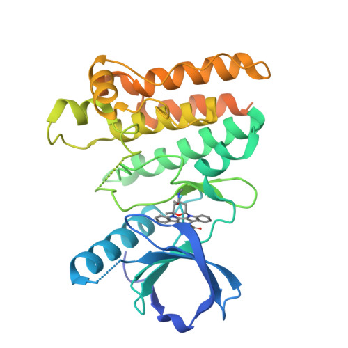
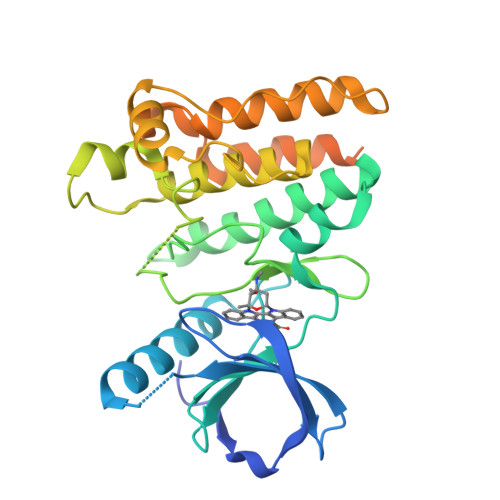
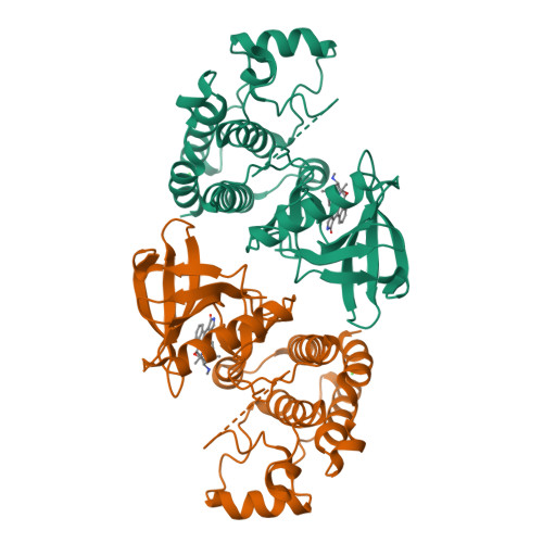
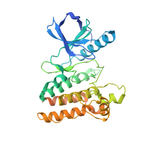
Crystal structure of murine receptor-interacting protein 4 (Ripk4) D143N bound to staurosporine


Experimental Data Snapshot

  • Method: X-RAY DIFFRACTION
  • Resolution: 2.50 Å
  • R-Value Free: 0.256 
  • R-Value Work: 0.223 
  • R-Value Observed: 0.224 

wwPDB Validation   3D Report Full Report


Ligand Structure Quality Assessment 


This is version 1.2 of the entry. See complete history


Literature

Crystal Structure of Ripk4 Reveals Dimerization-Dependent Kinase Activity.

Huang, C.S.Oberbeck, N.Hsiao, Y.C.Liu, P.Johnson, A.R.Dixit, V.M.Hymowitz, S.G.

(2018) Structure 26: 767

  • DOI: https://doi.org/10.1016/j.str.2018.04.002
  • Primary Citation of Related Structures:  
    5WNI, 5WNJ, 5WNK, 5WNL, 5WNM

  • PubMed Abstract: 

    Receptor-interacting protein kinase 4 (RIPK4) is a highly conserved regulator of epidermal differentiation. Members of the RIPK family possess a common kinase domain as well as unique accessory domains that likely dictate subcellular localization and substrate preferences. Mutations in human RIPK4 manifest as Bartsocas-Papas syndrome (BPS), a genetic disorder characterized by severe craniofacial and limb abnormalities. We describe the structure of the murine Ripk4 (MmRipk4) kinase domain, in ATP- and inhibitor-bound forms. The crystallographic dimer of MmRipk4 is similar to those of RIPK2 and BRAF, and we show that the intact dimeric entity is required for MmRipk4 catalytic activity through a series of engineered mutations and cell-based assays. We also assess the impact of BPS mutations on protein structure and activity to elucidate the molecular origins of the disease.


  • Organizational Affiliation

    Department of Structural Biology, Genentech, Inc., 1 DNA Way, South San Francisco, CA 94080, USA.


Macromolecules
Find similar proteins by:  (by identity cutoff)  |  3D Structure
Entity ID: 1
MoleculeChains Sequence LengthOrganismDetailsImage
Receptor-interacting serine/threonine-protein kinase 4342Mus musculusMutation(s): 1 
Gene Names: Ripk4Ankrd3Pkk
EC: 2.7.11.1
UniProt
Find proteins for Q9ERK0 (Mus musculus)
Explore Q9ERK0 
Go to UniProtKB:  Q9ERK0
Entity Groups  
Sequence Clusters30% Identity50% Identity70% Identity90% Identity95% Identity100% Identity
UniProt GroupQ9ERK0
Sequence Annotations
Expand
  • Reference Sequence
Small Molecules
Ligands 2 Unique
IDChains Name / Formula / InChI Key2D Diagram3D Interactions
STU
Query on STU

Download Ideal Coordinates CCD File 
B [auth A]STAUROSPORINE
C28 H26 N4 O3
HKSZLNNOFSGOKW-FYTWVXJKSA-N
CL
Query on CL

Download Ideal Coordinates CCD File 
C [auth A]CHLORIDE ION
Cl
VEXZGXHMUGYJMC-UHFFFAOYSA-M
Experimental Data & Validation

Experimental Data

  • Method: X-RAY DIFFRACTION
  • Resolution: 2.50 Å
  • R-Value Free: 0.256 
  • R-Value Work: 0.223 
  • R-Value Observed: 0.224 
  • Space Group: I 21 21 21
Unit Cell:
Length ( Å )Angle ( ˚ )
a = 70.158α = 90
b = 109.563β = 90
c = 146.221γ = 90
Software Package:
Software NamePurpose
REFMACrefinement
HKL-2000data reduction
HKL-2000data scaling
PHASERphasing

Structure Validation

View Full Validation Report


Ligand Structure Quality Assessment 


Entry History 

Deposition Data

Revision History  (Full details and data files)

  • Version 1.0: 2018-05-09
    Type: Initial release
  • Version 1.1: 2018-05-16
    Changes: Data collection, Database references
  • Version 1.2: 2024-03-13
    Changes: Data collection, Database references