5WNK | pdb_00005wnk

Crystal structure of murine receptor-interacting protein 4 (Ripk4) D143N bound to TG100-115


Experimental Data Snapshot

  • Method: X-RAY DIFFRACTION
  • Resolution: 3.11 Å
  • R-Value Free: 
    0.272 (Depositor), 0.264 (DCC) 
  • R-Value Work: 
    0.225 (Depositor), 0.225 (DCC) 
  • R-Value Observed: 
    0.228 (Depositor) 

wwPDB Validation 3D Report Full Report

Validation slider image for 5WNK

Ligand Structure Quality Assessment 


This is version 1.2 of the entry. See complete history

Literature

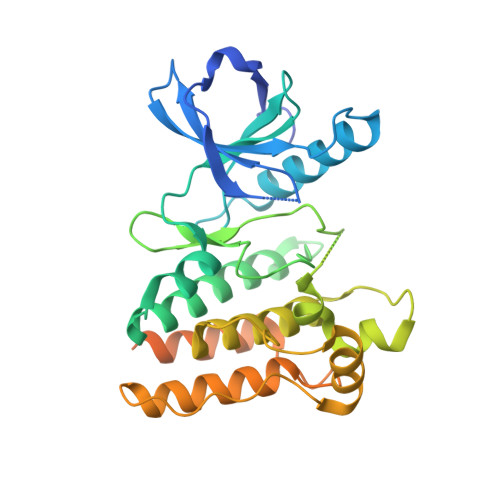
Crystal Structure of Ripk4 Reveals Dimerization-Dependent Kinase Activity.

Huang, C.S.Oberbeck, N.Hsiao, Y.C.Liu, P.Johnson, A.R.Dixit, V.M.Hymowitz, S.G.

(2018) Structure 26: 767

  • DOI: https://doi.org/10.1016/j.str.2018.04.002
  • Primary Citation Related Structures: 
    5WNI, 5WNJ, 5WNK, 5WNL, 5WNM

  • PubMed Abstract: 

    Receptor-interacting protein kinase 4 (RIPK4) is a highly conserved regulator of epidermal differentiation. Members of the RIPK family possess a common kinase domain as well as unique accessory domains that likely dictate subcellular localization and substrate preferences. Mutations in human RIPK4 manifest as Bartsocas-Papas syndrome (BPS), a genetic disorder characterized by severe craniofacial and limb abnormalities. We describe the structure of the murine Ripk4 (MmRipk4) kinase domain, in ATP- and inhibitor-bound forms. The crystallographic dimer of MmRipk4 is similar to those of RIPK2 and BRAF, and we show that the intact dimeric entity is required for MmRipk4 catalytic activity through a series of engineered mutations and cell-based assays. We also assess the impact of BPS mutations on protein structure and activity to elucidate the molecular origins of the disease.


  • Organizational Affiliation
    • Department of Structural Biology, Genentech, Inc., 1 DNA Way, South San Francisco, CA 94080, USA.

Macromolecule Content 

  • Total Structure Weight: 39.37 kDa 
  • Atom Count: 2,035 
  • Modeled Residue Count: 263 
  • Deposited Residue Count: 342 
  • Unique protein chains: 1

Macromolecules

Find similar proteins by:|  3D Structure
Entity ID: 1
MoleculeChains  Sequence LengthOrganismDetailsImage
Receptor-interacting serine/threonine-protein kinase 4342Mus musculusMutation(s): 1 
Gene Names: Ripk4Ankrd3Pkk
EC: 2.7.11.1
UniProt
Find proteins for Q9ERK0 (Mus musculus)
Explore Q9ERK0 
Go to UniProtKB:  Q9ERK0
Entity Groups
Sequence Clusters30% Identity50% Identity70% Identity90% Identity95% Identity100% Identity
UniProt GroupQ9ERK0
Sequence Annotations
Expand
Reference Sequence

Small Molecules

Ligands 2 Unique
IDChains Name / Formula / InChI Key2D Diagram3D Interactions
B6J

Query on B6J


Download:Ideal Coordinates CCD File
C [auth A]3,3'-(2,4-diaminopteridine-6,7-diyl)diphenol
C18 H14 N6 O2
UJIAQDJKSXQLIT-UHFFFAOYSA-N
CL

Query on CL


Download:Ideal Coordinates CCD File
B [auth A]CHLORIDE ION
Cl
VEXZGXHMUGYJMC-UHFFFAOYSA-M

Experimental Data & Validation

Experimental Data

  • Method: X-RAY DIFFRACTION
  • Resolution: 3.11 Å
  • R-Value Free:  0.272 (Depositor), 0.264 (DCC) 
  • R-Value Work:  0.225 (Depositor), 0.225 (DCC) 
  • R-Value Observed: 0.228 (Depositor) 
Space Group: I 21 21 21
Unit Cell:
Length ( Å )Angle ( ˚ )
a = 69.769α = 90
b = 110.231β = 90
c = 146.277γ = 90
Software Package:
Software NamePurpose
REFMACrefinement
HKL-2000data reduction
HKL-2000data scaling
PHASERphasing

Structure Validation

View Full Validation Report


Ligand Structure Quality Assessment 


Entry History 

Deposition Data

Revision History  (Full details and data files)

  • Version 1.0: 2018-05-09
    Type: Initial release
  • Version 1.1: 2018-05-16
    Changes: Data collection, Database references
  • Version 1.2: 2024-03-13
    Changes: Data collection, Database references