5WKI | pdb_00005wki

Crystal structure of PG90 TCR-CD1b-PG complex


Experimental Data Snapshot

  • Method: X-RAY DIFFRACTION
  • Resolution: 2.75 Å
  • R-Value Free: 
    0.252 (Depositor), 0.269 (DCC) 
  • R-Value Work: 
    0.212 (Depositor), 0.223 (DCC) 
  • R-Value Observed: 
    0.214 (Depositor) 

wwPDB Validation 3D Report Full Report

Validation slider image for 5WKI

Ligand Structure Quality Assessment 


This is version 2.1 of the entry. See complete history

Literature

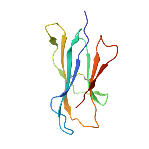
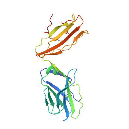
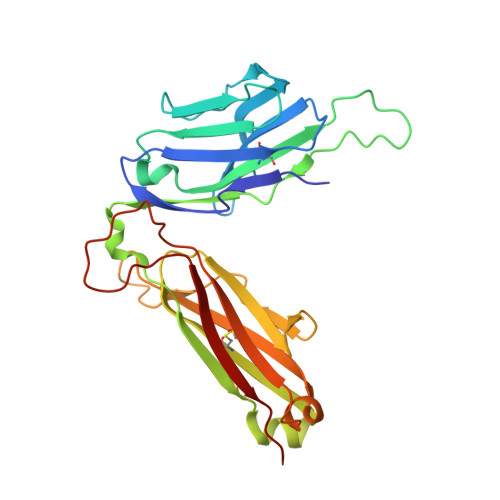
A molecular basis of human T cell receptor autoreactivity toward self-phospholipids.

Shahine, A.Van Rhijn, I.Cheng, T.Y.Iwany, S.Gras, S.Moody, D.B.Rossjohn, J.

(2017) Sci Immunol 2

  • DOI: https://doi.org/10.1126/sciimmunol.aao1384
  • Primary Citation Related Structures: 
    5WJO, 5WKE, 5WKG, 5WKI, 5WL1

  • PubMed Abstract: 

    Human T cell autoreactivity toward lipid antigens presented by CD1 proteins can manifest in numerous diseases, including psoriasis, contact hypersensitivities, and allergies. However, the molecular mechanisms for regulating T cell autoreactivity toward lipid antigens remain unclear. We determined the basis for T cell receptor (TCR) autoreactivity toward CD1b bound to self-phospholipids. The spectrum of self-antigens captured by CD1b skews toward abundant membrane phospholipids such as phosphatidylcholine and phosphatidylethanolamine. However, TCRs can specifically recognize rare phospholipids, including phosphatidylglycerol (PG). The structure of an autoreactive TCR bound to CD1b-PG shows that discrimination occurs through a marked induced fit movement of PG so that its polar head group fits snugly into the cationic cup of the TCR. Conversely, TCR binding toward ubiquitous self-phospholipids was sterically or electrostatically repelled. Accordingly, we describe a mechanism of TCR autoreactivity toward rare phospholipids and avoidance of autoreactivity to the most abundant self-phospholipids.


  • Organizational Affiliation
    • Infection and Immunity Program and Department of Biochemistry and Molecular Biology, Biomedicine Discovery Institute, Monash University, Clayton, Victoria 3800, Australia.

Macromolecule Content 

  • Total Structure Weight: 99.16 kDa 
  • Atom Count: 6,732 
  • Modeled Residue Count: 810 
  • Deposited Residue Count: 852 
  • Unique protein chains: 4

Macromolecules

Find similar proteins by:|  3D Structure
Entity ID: 1
MoleculeChains  Sequence LengthOrganismDetailsImage
T-cell surface glycoprotein CD1b300Homo sapiensMutation(s): 0 
Gene Names: CD1B
UniProt & NIH Common Fund Data Resources
Find proteins for P29016 (Homo sapiens)
Explore P29016 
Go to UniProtKB:  P29016
PHAROS:  P29016
GTEx:  ENSG00000158485 
Entity Groups
Sequence Clusters30% Identity50% Identity70% Identity90% Identity95% Identity100% Identity
UniProt GroupP29016
Glycosylation
Glycosylation Sites: 2Go to GlyGen: P29016-1
Sequence Annotations
Expand
Reference Sequence
Find similar proteins by:|  3D Structure
Entity ID: 2
MoleculeChains  Sequence LengthOrganismDetailsImage
Beta-2-microglobulin99Homo sapiensMutation(s): 0 
Gene Names: B2MCDABP0092HDCMA22P
UniProt & NIH Common Fund Data Resources
Find proteins for P61769 (Homo sapiens)
Explore P61769 
Go to UniProtKB:  P61769
PHAROS:  P61769
GTEx:  ENSG00000166710 
Entity Groups
Sequence Clusters30% Identity50% Identity70% Identity90% Identity95% Identity100% Identity
UniProt GroupP61769
Sequence Annotations
Expand
Reference Sequence
Find similar proteins by:|  3D Structure
Entity ID: 3
MoleculeChains  Sequence LengthOrganismDetailsImage
T-cell receptor alpha variable 26-1,TRA@ proteinC [auth D]204Homo sapiensMutation(s): 0 
Gene Names: TRAV26-1TRA@
UniProt & NIH Common Fund Data Resources
Find proteins for A0A087WT03 (Homo sapiens)
Explore A0A087WT03 
Go to UniProtKB:  A0A087WT03
PHAROS:  A0A087WT03
GTEx:  ENSG00000211807 
Find proteins for Q6P4G7 (Homo sapiens)
Explore Q6P4G7 
Go to UniProtKB:  Q6P4G7
Entity Groups
Sequence Clusters30% Identity50% Identity70% Identity90% Identity95% Identity100% Identity
UniProt GroupsA0A087WT03Q6P4G7
Sequence Annotations
Expand
Reference Sequence
Find similar proteins by:|  3D Structure
Entity ID: 4
MoleculeChains  Sequence LengthOrganismDetailsImage
PG90 TCR beta chainD [auth E]249Homo sapiensMutation(s): 0 

Oligosaccharides

Help  
Entity ID: 5
MoleculeChains Length2D Diagram GlycosylationD Interactions
alpha-L-fucopyranose-(1-3)-[2-acetamido-2-deoxy-beta-D-glucopyranose-(1-4)][alpha-L-fucopyranose-(1-6)]2-acetamido-2-deoxy-beta-D-glucopyranoseE [auth C]4N-Glycosylation
Glycosylation Resources
GlyTouCan: G63564LA
GlyCosmos: G63564LA
GlyGen: G63564LA

Small Molecules

Ligands 7 Unique
IDChains Name / Formula / InChI Key2D Diagram3D Interactions
D3D

Query on D3D


Download:Ideal Coordinates CCD File
U [auth D](19S,22R,25R)-22,25,26-trihydroxy-16,22-dioxo-17,21,23-trioxa-22lambda~5~-phosphahexacosan-19-yl (9E)-octadec-9-enoate
C40 H77 O10 P
PAZGBAOHGQRCBP-UCZVXENKSA-N
CUY

Query on CUY


Download:Ideal Coordinates CCD File
F [auth A]tetracosyl octadecanoate
C42 H84 O2
JKKQRJTZBKHHOI-UHFFFAOYSA-N
NAG

Query on NAG


Download:Ideal Coordinates CCD File
G [auth A]2-acetamido-2-deoxy-beta-D-glucopyranose
C8 H15 N O6
OVRNDRQMDRJTHS-FMDGEEDCSA-N
EDO

Query on EDO


Download:Ideal Coordinates CCD File
H [auth A]
I [auth A]
J [auth A]
K [auth A]
L [auth A]
H [auth A],
I [auth A],
J [auth A],
K [auth A],
L [auth A],
M [auth B],
N [auth D],
O [auth D],
P [auth D],
Q [auth D],
R [auth D],
S [auth D]
1,2-ETHANEDIOL
C2 H6 O2
LYCAIKOWRPUZTN-UHFFFAOYSA-N
ACT

Query on ACT


Download:Ideal Coordinates CCD File
V [auth E],
W [auth E]
ACETATE ION
C2 H3 O2
QTBSBXVTEAMEQO-UHFFFAOYSA-M
CL

Query on CL


Download:Ideal Coordinates CCD File
Y [auth E]CHLORIDE ION
Cl
VEXZGXHMUGYJMC-UHFFFAOYSA-M
NA

Query on NA


Download:Ideal Coordinates CCD File
T [auth D],
X [auth E]
SODIUM ION
Na
FKNQFGJONOIPTF-UHFFFAOYSA-N

Experimental Data & Validation

Experimental Data

  • Method: X-RAY DIFFRACTION
  • Resolution: 2.75 Å
  • R-Value Free:  0.252 (Depositor), 0.269 (DCC) 
  • R-Value Work:  0.212 (Depositor), 0.223 (DCC) 
  • R-Value Observed: 0.214 (Depositor) 
Space Group: C 1 2 1
Unit Cell:
Length ( Å )Angle ( ˚ )
a = 152.01α = 90
b = 82.97β = 94.88
c = 90.16γ = 90
Software Package:
Software NamePurpose
BUSTERrefinement
iMOSFLMdata reduction
Aimlessdata scaling
PHASERphasing

Structure Validation

View Full Validation Report


Ligand Structure Quality Assessment 


Entry History 

Deposition Data

Revision History  (Full details and data files)

  • Version 1.0: 2017-11-01
    Type: Initial release
  • Version 2.0: 2020-07-29
    Type: Remediation
    Reason: Carbohydrate remediation
    Changes: Advisory, Atomic model, Data collection, Derived calculations, Structure summary
  • Version 2.1: 2024-11-20
    Changes: Advisory, Data collection, Database references, Derived calculations, Structure summary