5WBB | pdb_00005wbb

Peroxide Activation Regulated by Hydrogen Bonds within Artificial Cu Proteins - S112A


Experimental Data Snapshot

  • Method: X-RAY DIFFRACTION
  • Resolution: 1.50 Å
  • R-Value Free: 
    0.188 (Depositor), 0.199 (DCC) 
  • R-Value Work: 
    0.156 (Depositor), 0.166 (DCC) 
  • R-Value Observed: 
    0.158 (Depositor) 

Starting Model: experimental
View more details

wwPDB Validation 3D Report Full Report

Validation slider image for 5WBB

Ligand Structure Quality Assessment 


This is version 1.2 of the entry. See complete history

Literature

Peroxide Activation Regulated by Hydrogen Bonds within Artificial Cu Proteins.

Mann, S.I.Heinisch, T.Ward, T.R.Borovik, A.S.

(2017) J Am Chem Soc 139: 17289-17292

  • DOI: https://doi.org/10.1021/jacs.7b10452
  • Primary Citation Related Structures: 
    5WBA, 5WBB, 5WBD, 6ANX

  • PubMed Abstract: 

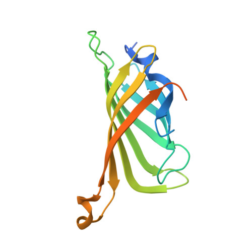
    Copper-hydroperoxido species (Cu II -OOH) have been proposed to be key intermediates in biological and synthetic oxidations. Using biotin-streptavidin (Sav) technology, artificial copper proteins have been developed to stabilize a Cu II -OOH complex in solution and in crystallo. Stability is achieved because the Sav host provides a local environment around the Cu-OOH that includes a network of hydrogen bonds to the hydroperoxido ligand. Systematic deletions of individual hydrogen bonds to the Cu-OOH complex were accomplished using different Sav variants and demonstrated that stability is achieved with a single hydrogen bond to the proximal O-atom of the hydroperoxido ligand: changing this interaction to only include the distal O-atom produced a reactive variant that oxidized an external substrate.


  • Organizational Affiliation
    • Department of Chemistry, University of California , 1102 Natural Science II, Irvine, California 92697, United States.

Macromolecule Content 

  • Total Structure Weight: 17.37 kDa 
  • Atom Count: 1,128 
  • Modeled Residue Count: 125 
  • Deposited Residue Count: 159 
  • Unique protein chains: 1

Macromolecules

Find similar proteins by:|  3D Structure
Entity ID: 1
MoleculeChains  Sequence LengthOrganismDetailsImage
Streptavidin159Streptomyces avidiniiMutation(s): 1 
UniProt
Find proteins for P22629 (Streptomyces avidinii)
Explore P22629 
Go to UniProtKB:  P22629
Entity Groups
Sequence Clusters30% Identity50% Identity70% Identity90% Identity95% Identity100% Identity
UniProt GroupP22629
Sequence Annotations
Expand
Reference Sequence

Experimental Data & Validation

Experimental Data

  • Method: X-RAY DIFFRACTION
  • Resolution: 1.50 Å
  • R-Value Free:  0.188 (Depositor), 0.199 (DCC) 
  • R-Value Work:  0.156 (Depositor), 0.166 (DCC) 
  • R-Value Observed: 0.158 (Depositor) 
Space Group: I 41 2 2
Unit Cell:
Length ( Å )Angle ( ˚ )
a = 57.64α = 90
b = 57.64β = 90
c = 183.98γ = 90
Software Package:
Software NamePurpose
REFMACrefinement
iMOSFLMdata reduction
Aimlessdata scaling
PHASERphasing

Structure Validation

View Full Validation Report


Ligand Structure Quality Assessment 


Entry History 

Deposition Data

Revision History  (Full details and data files)

  • Version 1.0: 2017-11-22
    Type: Initial release
  • Version 1.1: 2017-12-13
    Changes: Database references
  • Version 1.2: 2023-10-04
    Changes: Data collection, Database references, Refinement description