5UHI | pdb_00005uhi

Structure of RORgt bound to


Experimental Data Snapshot

  • Method: X-RAY DIFFRACTION
  • Resolution: 3.20 Å
  • R-Value Free: 
    0.318 (Depositor), 0.284 (DCC) 
  • R-Value Work: 
    0.257 (Depositor) 
  • R-Value Observed: 
    0.263 (Depositor) 

wwPDB Validation 3D Report Full Report

Validation slider image for 5UHI

Ligand Structure Quality Assessment 


This is version 1.2 of the entry. See complete history

Literature

Identification and structure activity relationships of quinoline tertiary alcohol modulators of ROR gamma t.

Kummer, D.A.Cummings, M.D.Abad, M.Barbay, J.Castro, G.Wolin, R.Kreutter, K.D.Maharoof, U.Milligan, C.Nishimura, R.Pierce, J.Schalk-Hihi, C.Spurlino, J.Urbanski, M.Venkatesan, H.Wang, A.Woods, C.Xue, X.Edwards, J.P.Fourie, A.M.Leonard, K.

(2017) Bioorg Med Chem Lett 27: 2047-2057

  • DOI: https://doi.org/10.1016/j.bmcl.2017.02.044
  • Primary Citation Related Structures: 
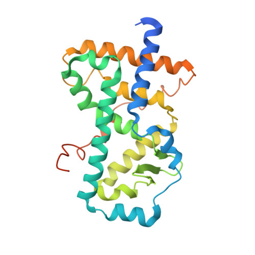
    5UFO, 5UFR, 5UHI

  • PubMed Abstract: 

    A high-throughput screen of the ligand binding domain of the nuclear receptor retinoic acid-related orphan receptor gamma t (RORγt) employing a thermal shift assay yielded a quinoline tertiary alcohol hit. Optimization of the 2-, 3- and 4-positions of the quinoline core using structure-activity relationships and structure-based drug design methods led to the discovery of a series of modulators with improved RORγt inhibitory potency and inverse agonism properties.


  • Organizational Affiliation
    • Discovery Immunology, Janssen Research and Development, 3210 Merryfield Row, San Diego, CA 92121, United States. Electronic address: dkummer1@its.jnj.com.

Macromolecule Content 

  • Total Structure Weight: 61.35 kDa 
  • Atom Count: 3,708 
  • Modeled Residue Count: 446 
  • Deposited Residue Count: 516 
  • Unique protein chains: 1

Macromolecules

Find similar proteins by:|  3D Structure
Entity ID: 1
MoleculeChains  Sequence LengthOrganismDetailsImage
Nuclear receptor ROR-gamma
A, B
258Homo sapiensMutation(s): 0 
Gene Names: RORCNR1F3RORGRZRG
UniProt & NIH Common Fund Data Resources
Find proteins for P51449 (Homo sapiens)
Explore P51449 
Go to UniProtKB:  P51449
PHAROS:  P51449
GTEx:  ENSG00000143365 
Entity Groups
Sequence Clusters30% Identity50% Identity70% Identity90% Identity95% Identity100% Identity
UniProt GroupP51449
Sequence Annotations
Expand
Reference Sequence

Small Molecules

Ligands 1 Unique
IDChains Name / Formula / InChI Key2D Diagram3D Interactions
8A4

Query on 8A4


Download:Ideal Coordinates CCD File
C [auth A],
D [auth B]
(R)-(4-chloro-2-methoxy-3-{[4-(1H-pyrazol-1-yl)phenyl]methyl}quinolin-6-yl)(4-chlorophenyl)(1-methyl-1H-imidazol-5-yl)methanol
C31 H25 Cl2 N5 O2
UWOBQISTECYGOP-WJOKGBTCSA-N

Experimental Data & Validation

Experimental Data

  • Method: X-RAY DIFFRACTION
  • Resolution: 3.20 Å
  • R-Value Free:  0.318 (Depositor), 0.284 (DCC) 
  • R-Value Work:  0.257 (Depositor) 
  • R-Value Observed: 0.263 (Depositor) 
Space Group: C 2 2 21
Unit Cell:
Length ( Å )Angle ( ˚ )
a = 104.35α = 90
b = 104.323β = 90
c = 123.577γ = 90
Software Package:
Software NamePurpose
PHENIXrefinement
HKL-2000data reduction
HKL-2000data scaling
PHENIXphasing

Structure Validation

View Full Validation Report


Ligand Structure Quality Assessment 


Entry History 

Deposition Data

Revision History  (Full details and data files)

  • Version 1.0: 2017-04-05
    Type: Initial release
  • Version 1.1: 2017-04-26
    Changes: Database references
  • Version 1.2: 2024-03-06
    Changes: Data collection, Database references