5TTF

Crystal structure of catalytic domain of G9a with MS012


Experimental Data Snapshot

  • Method: X-RAY DIFFRACTION
  • Resolution: 1.72 Å
  • R-Value Free: 0.247 
  • R-Value Work: 0.207 
  • R-Value Observed: 0.207 

Starting Model: experimental
View more details

wwPDB Validation   3D Report Full Report


Ligand Structure Quality Assessment 


This is version 1.2 of the entry. See complete history


Literature

Discovery of Potent and Selective Inhibitors for G9a-Like Protein (GLP) Lysine Methyltransferase.

Xiong, Y.Li, F.Babault, N.Dong, A.Zeng, H.Wu, H.Chen, X.Arrowsmith, C.H.Brown, P.J.Liu, J.Vedadi, M.Jin, J.

(2017) J Med Chem 60: 1876-1891

  • DOI: https://doi.org/10.1021/acs.jmedchem.6b01645
  • Primary Citation of Related Structures:  
    5TTF, 5TTG, 5TUY, 5TUZ

  • PubMed Abstract: 

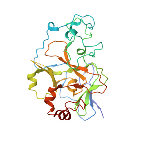
    G9a-like protein (GLP) and G9a are highly homologous protein lysine methyltransferases (PKMTs) sharing approximately 80% sequence identity in their catalytic domains. GLP and G9a form a heterodimer complex and catalyze mono- and dimethylation of histone H3 lysine 9 and nonhistone substrates. Although they are closely related, GLP and G9a possess distinct physiological and pathophysiological functions. Thus, GLP or G9a selective small-molecule inhibitors are useful tools to dissect their distinct biological functions. We previously reported potent and selective G9a/GLP dual inhibitors including UNC0638 and UNC0642. Here we report the discovery of potent and selective GLP inhibitors including 4 (MS0124) and 18 (MS012), which are >30-fold and 140-fold selective for GLP over G9a and other methyltransferases, respectively. The cocrystal structures of GLP and G9a in the complex with either 4 or 18 displayed virtually identical binding modes and interactions, highlighting the challenges in structure-based design of selective inhibitors for either enzyme.


  • Organizational Affiliation

    Department of Pharmacological Sciences and Department of Oncological Sciences, Icahn School of Medicine at Mount Sinai , New York, New York 10029, United States.


Macromolecules
Find similar proteins by:  (by identity cutoff)  |  3D Structure
Entity ID: 1
MoleculeChains Sequence LengthOrganismDetailsImage
Histone-lysine N-methyltransferase EHMT2
A, B, C, D
283Homo sapiensMutation(s): 0 
Gene Names: EHMT2BAT8C6orf30G9AKMT1CNG36
EC: 2.1.1 (PDB Primary Data), 2.1.1.43 (PDB Primary Data)
UniProt & NIH Common Fund Data Resources
Find proteins for Q96KQ7 (Homo sapiens)
Explore Q96KQ7 
Go to UniProtKB:  Q96KQ7
GTEx:  ENSG00000204371 
Entity Groups  
Sequence Clusters30% Identity50% Identity70% Identity90% Identity95% Identity100% Identity
UniProt GroupQ96KQ7
Sequence Annotations
Expand
  • Reference Sequence
Small Molecules
Ligands 5 Unique
IDChains Name / Formula / InChI Key2D Diagram3D Interactions
7KZ
Query on 7KZ

Download Ideal Coordinates CCD File 
GA [auth D],
J [auth A],
R [auth B],
Z [auth C]
N4-(1-methylpiperidin-4-yl)-N2-hexyl-6,7-dimethoxyquinazoline-2,4-diamine
C22 H35 N5 O2
ZYNUWSFRZCRKSN-UHFFFAOYSA-N
SAM
Query on SAM

Download Ideal Coordinates CCD File 
FA [auth D],
I [auth A],
Q [auth B],
Y [auth C]
S-ADENOSYLMETHIONINE
C15 H22 N6 O5 S
MEFKEPWMEQBLKI-FCKMPRQPSA-N
ZN
Query on ZN

Download Ideal Coordinates CCD File 
BA [auth D]
CA [auth D]
DA [auth D]
E [auth A]
EA [auth D]
BA [auth D],
CA [auth D],
DA [auth D],
E [auth A],
EA [auth D],
F [auth A],
G [auth A],
H [auth A],
M [auth B],
N [auth B],
O [auth B],
P [auth B],
U [auth C],
V [auth C],
W [auth C],
X [auth C]
ZINC ION
Zn
PTFCDOFLOPIGGS-UHFFFAOYSA-N
CL
Query on CL

Download Ideal Coordinates CCD File 
K [auth A]CHLORIDE ION
Cl
VEXZGXHMUGYJMC-UHFFFAOYSA-M
UNX
Query on UNX

Download Ideal Coordinates CCD File 
AA [auth C],
HA [auth D],
L [auth A],
S [auth B],
T [auth B]
UNKNOWN ATOM OR ION
X
Binding Affinity Annotations 
IDSourceBinding Affinity
7KZ BindingDB:  5TTF Kd: 610 (nM) from 1 assay(s)
IC50: 992 (nM) from 1 assay(s)
Experimental Data & Validation

Experimental Data

  • Method: X-RAY DIFFRACTION
  • Resolution: 1.72 Å
  • R-Value Free: 0.247 
  • R-Value Work: 0.207 
  • R-Value Observed: 0.207 
  • Space Group: P 1
  • Diffraction Data: https://doi.org/10.18430/M35TTF
Unit Cell:
Length ( Å )Angle ( ˚ )
a = 56.846α = 92.09
b = 67.749β = 90.05
c = 77.845γ = 91.8
Software Package:
Software NamePurpose
REFMACrefinement
SCALEPACKdata scaling
PDB_EXTRACTdata extraction
PHASERphasing
HKL-3000data reduction

Structure Validation

View Full Validation Report


Ligand Structure Quality Assessment 


Entry History 

Revision History  (Full details and data files)

  • Version 1.0: 2016-12-21
    Type: Initial release
  • Version 1.1: 2018-05-23
    Changes: Data collection, Database references
  • Version 1.2: 2023-10-04
    Changes: Data collection, Database references, Derived calculations, Refinement description