5TGD | pdb_00005tgd

Crystal structure of FolM Alternative dihydrofolate reductase 1 from Brucella suis in complex with NADP


Experimental Data Snapshot

  • Method: X-RAY DIFFRACTION
  • Resolution: 1.70 Å
  • R-Value Free: 
    0.198 (Depositor), 0.198 (DCC) 
  • R-Value Work: 
    0.163 (Depositor), 0.164 (DCC) 
  • R-Value Observed: 
    0.163 (Depositor) 

Starting Model: experimental
View more details

wwPDB Validation 3D Report Full Report

Validation slider image for 5TGD

Ligand Structure Quality Assessment 


This is version 1.2 of the entry. See complete history

Literature

Crystal structures of FolM alternative dihydrofolate reductase 1 from Brucella suis and Brucella canis.

Porter, I.Neal, T.Walker, Z.Hayes, D.Fowler, K.Billups, N.Rhoades, A.Smith, C.Smith, K.Staker, B.L.Dranow, D.M.Mayclin, S.J.Subramanian, S.Edwards, T.E.Myler, P.J.Asojo, O.A.

(2022) Acta Crystallogr F Struct Biol Commun 78: 31-38

  • DOI: https://doi.org/10.1107/S2053230X21013078
  • Primary Citation Related Structures: 
    5BT9, 5TGD

  • PubMed Abstract: 

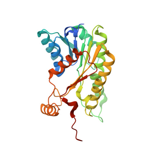
    Members of the bacterial genus Brucella cause brucellosis, a zoonotic disease that affects both livestock and wildlife. Brucella are category B infectious agents that can be aerosolized for biological warfare. As part of the structural genomics studies at the Seattle Structural Genomics Center for Infectious Disease (SSGCID), FolM alternative dihydrofolate reductases 1 from Brucella suis and Brucella canis were produced and their structures are reported. The enzymes share ∼95% sequence identity but have less than 33% sequence identity to other homologues with known structure. The structures are prototypical NADPH-dependent short-chain reductases that share their highest tertiary-structural similarity with protozoan pteridine reductases, which are being investigated for rational therapeutic development.


  • Organizational Affiliation
    • Department of Chemistry and Biochemistry, Hampton University, 100 William R. Harvey Way, Hampton, VA 23668, USA.

Macromolecule Content 

  • Total Structure Weight: 122.09 kDa 
  • Atom Count: 8,598 
  • Modeled Residue Count: 983 
  • Deposited Residue Count: 1,068 
  • Unique protein chains: 1

Macromolecules

Find similar proteins by:|  3D Structure
Entity ID: 1
MoleculeChains  Sequence LengthOrganismDetailsImage
FolM Alternative dihydrofolate reductase
A, B, C, D
267Brucella suis 92/29Mutation(s): 0 
Gene Names: C062_00524

Small Molecules

Ligands 3 Unique
IDChains Name / Formula / InChI Key2D Diagram3D Interactions
NAP

Query on NAP


Download:Ideal Coordinates CCD File
E [auth A],
H [auth B],
I [auth C],
K [auth D]
NADP NICOTINAMIDE-ADENINE-DINUCLEOTIDE PHOSPHATE
C21 H28 N7 O17 P3
XJLXINKUBYWONI-NNYOXOHSSA-N
GOL

Query on GOL


Download:Ideal Coordinates CCD File
G [auth A]GLYCEROL
C3 H8 O3
PEDCQBHIVMGVHV-UHFFFAOYSA-N
EDO

Query on EDO


Download:Ideal Coordinates CCD File
F [auth A],
J [auth C],
L [auth D],
M [auth D]
1,2-ETHANEDIOL
C2 H6 O2
LYCAIKOWRPUZTN-UHFFFAOYSA-N

Experimental Data & Validation

Experimental Data

  • Method: X-RAY DIFFRACTION
  • Resolution: 1.70 Å
  • R-Value Free:  0.198 (Depositor), 0.198 (DCC) 
  • R-Value Work:  0.163 (Depositor), 0.164 (DCC) 
  • R-Value Observed: 0.163 (Depositor) 
Space Group: P 1 21 1
Unit Cell:
Length ( Å )Angle ( ˚ )
a = 76.35α = 90
b = 76.52β = 109.47
c = 98.26γ = 90
Software Package:
Software NamePurpose
XSCALEdata scaling
MOLREPphasing
PHENIXrefinement
PDB_EXTRACTdata extraction
Cootmodel building

Structure Validation

View Full Validation Report


Ligand Structure Quality Assessment 


Entry History 

Deposition Data

Revision History  (Full details and data files)

  • Version 1.0: 2016-10-12
    Type: Initial release
  • Version 1.1: 2023-09-20
    Changes: Data collection, Database references, Derived calculations
  • Version 1.2: 2023-10-04
    Changes: Refinement description