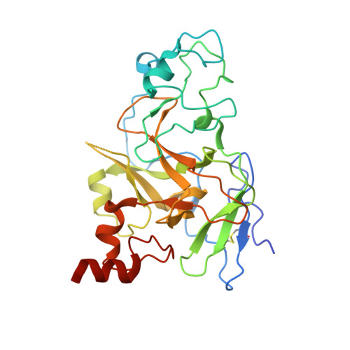
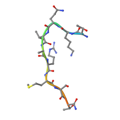
5T0K

Structure of G9a SET-domain with H3K9M mutant peptide and SAM

  • Classification: TRANSFERASE
  • Organism(s): Homo sapiens
  • Expression System: Escherichia coli
  • Mutation(s): No 

  • Deposited: 2016-08-16 Released: 2016-10-05 
  • Deposition Author(s): Xu, K., Tong, L.
  • Funding Organization(s): National Institutes of Health/National Institute of General Medical Sciences (NIH/NIGMS), National Institutes of Health/Office of the Director, National Institutes of Health/National Center for Research Resources (NIH/NCRR)

Experimental Data Snapshot

  • Method: X-RAY DIFFRACTION
  • Resolution: 1.70 Å
  • R-Value Free: 0.193 
  • R-Value Work: 0.165 
  • R-Value Observed: 0.166 

Starting Model: experimental
View more details

wwPDB Validation   3D Report Full Report


Ligand Structure Quality Assessment 


This is version 1.3 of the entry. See complete history


Literature

A histone H3K9M mutation traps histone methyltransferase Clr4 to prevent heterochromatin spreading.

Shan, C.M.Wang, J.Xu, K.Chen, H.Yue, J.X.Andrews, S.Moresco, J.J.Yates, J.R.Nagy, P.L.Tong, L.Jia, S.

(2016) Elife 5

  • DOI: https://doi.org/10.7554/eLife.17903
  • Primary Citation of Related Structures:  
    5T0K, 5T0M

  • PubMed Abstract: 

    Histone lysine-to-methionine (K-to-M) mutations are associated with multiple cancers, and they function in a dominant fashion to block the methylation of corresponding lysines on wild type histones. However, their mechanisms of function are controversial. Here we show that in fission yeast, introducing the K9M mutation into one of the three histone H3 genes dominantly blocks H3K9 methylation on wild type H3 across the genome. In addition, H3K9M enhances the interaction of histone H3 tail with the H3K9 methyltransferase Clr4 in a SAM (S-adenosyl-methionine)-dependent manner, and Clr4 is trapped at nucleation sites to prevent its spreading and the formation of large heterochromatin domains. We further determined the crystal structure of an H3K9M peptide in complex with human H3K9 methyltransferase G9a and SAM, which reveales that the methionine side chain had enhanced van der Waals interactions with G9a. Therefore, our results provide a detailed mechanism by which H3K9M regulates H3K9 methylation.


  • Organizational Affiliation

    Department of Biological Sciences, Columbia University, New York, United States.


Macromolecules
Find similar proteins by:  (by identity cutoff)  |  3D Structure
Entity ID: 1
MoleculeChains Sequence LengthOrganismDetailsImage
Histone-lysine N-methyltransferase EHMT2
A, B
281Homo sapiensMutation(s): 0 
Gene Names: EHMT2BAT8C6orf30G9AKMT1CNG36
EC: 2.1.1 (PDB Primary Data), 2.1.1.43 (PDB Primary Data), 2.1.1.367 (UniProt)
UniProt & NIH Common Fund Data Resources
Find proteins for Q96KQ7 (Homo sapiens)
Explore Q96KQ7 
Go to UniProtKB:  Q96KQ7
PHAROS:  Q96KQ7
GTEx:  ENSG00000204371 
Entity Groups  
Sequence Clusters30% Identity50% Identity70% Identity90% Identity95% Identity100% Identity
UniProt GroupQ96KQ7
Sequence Annotations
Expand
  • Reference Sequence

Find similar proteins by:  Sequence   |   3D Structure  

Entity ID: 2
MoleculeChains Sequence LengthOrganismDetailsImage
H3K9 mutant peptideC [auth P],
D [auth Q]
15Homo sapiensMutation(s): 0 
UniProt & NIH Common Fund Data Resources
Find proteins for P68431 (Homo sapiens)
Explore P68431 
Go to UniProtKB:  P68431
PHAROS:  P68431
Entity Groups  
Sequence Clusters30% Identity50% Identity70% Identity90% Identity95% Identity100% Identity
UniProt GroupP68431
Sequence Annotations
Expand
  • Reference Sequence
Small Molecules
Ligands 2 Unique
IDChains Name / Formula / InChI Key2D Diagram3D Interactions
SAM
Query on SAM

Download Ideal Coordinates CCD File 
I [auth A],
N [auth B]
S-ADENOSYLMETHIONINE
C15 H22 N6 O5 S
MEFKEPWMEQBLKI-FCKMPRQPSA-N
ZN
Query on ZN

Download Ideal Coordinates CCD File 
E [auth A]
F [auth A]
G [auth A]
H [auth A]
J [auth B]
E [auth A],
F [auth A],
G [auth A],
H [auth A],
J [auth B],
K [auth B],
L [auth B],
M [auth B]
ZINC ION
Zn
PTFCDOFLOPIGGS-UHFFFAOYSA-N
Experimental Data & Validation

Experimental Data

  • Method: X-RAY DIFFRACTION
  • Resolution: 1.70 Å
  • R-Value Free: 0.193 
  • R-Value Work: 0.165 
  • R-Value Observed: 0.166 
  • Space Group: P 21 21 21
Unit Cell:
Length ( Å )Angle ( ˚ )
a = 75.101α = 90
b = 92.307β = 90
c = 93.143γ = 90
Software Package:
Software NamePurpose
PHENIXrefinement
HKL-2000data reduction
HKL-2000data scaling
PHENIXphasing

Structure Validation

View Full Validation Report


Ligand Structure Quality Assessment 


Entry History & Funding Information

Deposition Data


Funding OrganizationLocationGrant Number
National Institutes of Health/National Institute of General Medical Sciences (NIH/NIGMS)United StatesR01-GM085145
National Institutes of Health/Office of the DirectorUnited StatesS10-OD012018
National Institutes of Health/National Center for Research Resources (NIH/NCRR)United StatesP41-RR011823
National Institutes of Health/National Institute of General Medical Sciences (NIH/NIGMS)United StatesP41-GM103533

Revision History  (Full details and data files)

  • Version 1.0: 2016-10-05
    Type: Initial release
  • Version 1.1: 2017-09-20
    Changes: Author supporting evidence, Derived calculations
  • Version 1.2: 2019-12-04
    Changes: Author supporting evidence
  • Version 1.3: 2023-10-04
    Changes: Data collection, Database references, Derived calculations, Refinement description