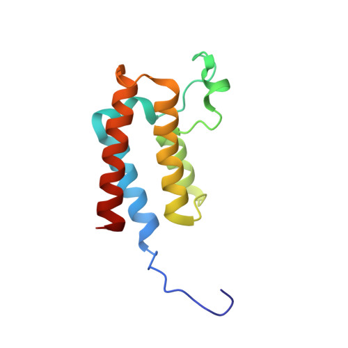
5ORB

Crystal Structure of BAZ2B bromodomain in complex with 1-methyl-cyclopentapyrazole compound 30


Experimental Data Snapshot

  • Method: X-RAY DIFFRACTION
  • Resolution: 2.10 Å
  • R-Value Free: 0.218 
  • R-Value Work: 0.188 
  • R-Value Observed: 0.189 

Starting Model: experimental
View more details

wwPDB Validation   3D Report Full Report


Ligand Structure Quality Assessment 


This is version 1.2 of the entry. See complete history


Literature

Discovery of Inhibitors of Four Bromodomains by Fragment-Anchored Ligand Docking.

Marchand, J.R.Dalle Vedove, A.Lolli, G.Caflisch, A.

(2017) J Chem Inf Model 57: 2584-2597

  • DOI: https://doi.org/10.1021/acs.jcim.7b00336
  • Primary Citation of Related Structures:  
    5OR8, 5OR9, 5ORB

  • PubMed Abstract: 

    The high-throughput docking protocol called ALTA-VS (anchor-based library tailoring approach for virtual screening) was developed in 2005 for the efficient in silico screening of large libraries of compounds by preselection of only those molecules that have optimal fragments (anchors) for the protein target. Here we present an updated version of ALTA-VS with a broader range of potential applications. The evaluation of binding energy makes use of a classical force field with implicit solvent in the continuum dielectric approximation. In about 2 days per protein target on a 96-core compute cluster (equipped with Xeon E3-1280 quad core processors at 2.5 GHz), the screening of a library of nearly 77 000 diverse molecules with the updated ALTA-VS protocol has resulted in the identification of 19, 3, 3, and 2 μM inhibitors of the human bromodomains ATAD2, BAZ2B, BRD4(1), and CREBBP, respectively. The success ratio (i.e., number of actives in a competition binding assay in vitro divided by the number of compounds tested) ranges from 8% to 13% in dose-response measurements. The poses predicted by fragment-based docking for the three ligands of the BAZ2B bromodomain were confirmed by protein X-ray crystallography.


  • Organizational Affiliation

    Department of Biochemistry, University of Zürich , CH-8057, Zürich, Switzerland.


Macromolecules
Find similar proteins by:  (by identity cutoff)  |  3D Structure
Entity ID: 1
MoleculeChains Sequence LengthOrganismDetailsImage
Bromodomain adjacent to zinc finger domain protein 2B117Homo sapiensMutation(s): 0 
Gene Names: BAZ2BKIAA1476
UniProt & NIH Common Fund Data Resources
Find proteins for Q9UIF8 (Homo sapiens)
Explore Q9UIF8 
Go to UniProtKB:  Q9UIF8
PHAROS:  Q9UIF8
GTEx:  ENSG00000123636 
Entity Groups  
Sequence Clusters30% Identity50% Identity70% Identity90% Identity95% Identity100% Identity
UniProt GroupQ9UIF8
Sequence Annotations
Expand
  • Reference Sequence
Small Molecules
Ligands 2 Unique
IDChains Name / Formula / InChI Key2D Diagram3D Interactions
JR6
Query on JR6

Download Ideal Coordinates CCD File 
B [auth A]2-(4-methoxyphenyl)sulfanyl-~{N}-(2-methyl-5,6-dihydro-4~{H}-cyclopenta[c]pyrazol-3-yl)ethanamide
C16 H19 N3 O2 S
HLIFAMDWHQBTKJ-UHFFFAOYSA-N
EDO
Query on EDO

Download Ideal Coordinates CCD File 
C [auth A]1,2-ETHANEDIOL
C2 H6 O2
LYCAIKOWRPUZTN-UHFFFAOYSA-N
Experimental Data & Validation

Experimental Data

  • Method: X-RAY DIFFRACTION
  • Resolution: 2.10 Å
  • R-Value Free: 0.218 
  • R-Value Work: 0.188 
  • R-Value Observed: 0.189 
  • Space Group: C 2 2 21
Unit Cell:
Length ( Å )Angle ( ˚ )
a = 80.906α = 90
b = 96.788β = 90
c = 57.82γ = 90
Software Package:
Software NamePurpose
PHASERphasing
PHENIXrefinement
PDB_EXTRACTdata extraction
XDSdata scaling
XDSdata reduction

Structure Validation

View Full Validation Report


Ligand Structure Quality Assessment 


Entry History & Funding Information

Deposition Data


Funding OrganizationLocationGrant Number
Swiss Cancer Society (Krebsliga Schweiz)SwitzerlandKLS-3098- 02-2013

Revision History  (Full details and data files)

  • Version 1.0: 2017-09-13
    Type: Initial release
  • Version 1.1: 2017-11-01
    Changes: Database references
  • Version 1.2: 2024-01-17
    Changes: Data collection, Database references, Refinement description